Blog
Blog
AI in Taiwan Radiology: Lunit, AIDOC, and What "AI-Assisted Screening" Actually Means
April 14, 2026
The phrase "AI-assisted radiology" gets used loosely. In Taiwan's preventive screening centers, it means something specific: FDA/TFDA-cleared deep-learning models that pre-flag findings on imaging before a board-certified radiologist reads the scan. The most-deployed vendors at our partner hospitals include Lunit (lung nodules and chest radiology), AIDOC (head CT triage), Annalise.ai (chest x-ray comprehensive), HeartFlow (coronary functional analysis), and locally-developed models from National Taiwan University Hospital's AI Research Center and Chang Gung Memorial Hospital. The mechanism isn't replacement — it's a second pair of eyes that never gets tired and never has a bad day.
This is a long read because the topic is widely misunderstood. We're going to be specific about which products carry which clearances, what the published peer-reviewed evidence actually showed (and didn't show), how these tools fit into a real radiologist's worklist via PACS, and why locally-trained Taiwan models matter for Asian and Asian-American patients in ways that imported Western models historically have not.
What "AI-assisted radiology" actually means in 2026
"AI in radiology" covers a wide spectrum, much of which is marketing rather than clinical deployment. Inside the imaging suite of a TFDA-regulated screening hospital, the regulated tools fall into three distinct categories — and the distinction matters because each has different evidence requirements, different workflow integration patterns, and different risks.
- CADe (Computer-Aided Detection) — Models that mark suspicious regions on an image: a possible nodule on a chest x-ray, a possible bleed on a head CT, a calcification on a mammogram. CADe does not classify what the finding is. It only says "look here." Lunit INSIGHT CXR, Riverain ClearRead CXR, and AIDOC's head CT triage are predominantly CADe.
- CADx (Computer-Aided Diagnosis) — Models that go further and characterize a finding: benign vs malignant probability, lesion type classification, BI-RADS-equivalent scoring. The regulatory bar is higher. NTU's liver lesion characterization model and Lunit INSIGHT MMG (which produces an Abnormality Score) sit closer to CADx.
- Triage and notification tools — Models whose primary job isn't detection per se but reordering the radiologist's worklist so time-critical findings (intracranial hemorrhage, pulmonary embolism, aortic dissection) get read first. AIDOC's stroke and bleed modules are the canonical example, FDA-cleared specifically as triage.
None of these are autonomous diagnostic systems. None of them author reports. None of them communicate with patients. All of them sit upstream of a board-certified human and produce output that a radiologist either confirms, dismisses, or contextualizes against patient history.
Why this matters for screening accuracy
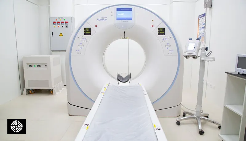
Preventive imaging produces large data volumes per patient — a full-body MRI generates ~3,000 images, a low-dose lung CT another ~400, a coronary CTA roughly 500 with multi-phase reconstruction. Subtle findings (a 4 mm pulmonary nodule, an early calcification, a small hepatic cyst, a 3 mm renal lesion) can be missed by even an experienced radiologist on a busy day with high case volume. Published evidence shows AI second-read tools improve sensitivity for early-stage lung nodule detection by roughly 15–20% (Lunit INSIGHT CXR, Lancet Digital Health 2020), reduce time-to-notification for acute intracranial hemorrhage by tens of minutes (AIDOC, NEJM AI 2023), and detect a clinically meaningful share of additional findings on chest x-rays that human readers missed (Annalise.ai, Radiology 2022).
The clinical workflow at our partner hospitals integrates AI in three places:
- Pre-read flagging: AI marks suspicious regions on the radiologist's worklist before review. The radiologist sees the AI's heat map alongside the original image and decides whether the flag is real.
- Quantitative measurement: AI auto-measures things — organ volume, lesion dimensions, coronary calcium scores in Agatston units, liver fat fraction, vertebral compression metrics — reducing measurement variance between readers and across visits.
- Comparison across visits: AI auto-aligns prior scans to current and highlights changes — critical for longitudinal monitoring of small nodules where 1 mm of growth over 12 months changes management.
Vendor-by-vendor — what's deployed at Taiwan partners
Below is the comprehensive table of AI tools deployed across our partner hospitals (NTU Hospital, Chang Gung Memorial, MacKay, Cathay General, NCKU). Sensitivity and specificity figures are from the cited peer-reviewed validation studies — they reflect performance under controlled conditions and are not a guarantee of real-world per-scan accuracy.
| Vendor / Product | Use case | Regulatory clearance | Reported performance |
|---|---|---|---|
| Lunit INSIGHT CXR | Chest x-ray — nodule, consolidation, pneumothorax, pleural effusion | FDA, TFDA, CE, KFDA | Sensitivity ~97% / Specificity ~90% (Lancet Digital Health 2020); ~15–20% sensitivity gain for early-stage lung nodules when used as second reader |
| Lunit INSIGHT MMG | Mammography — soft tissue lesions, calcifications | FDA, CE, MFDS | AUC 0.94–0.96 in published reader studies; non-inferior to a panel of breast radiologists |
| Lunit INSIGHT DBT | Digital breast tomosynthesis (3D mammography) | CE, KFDA | Reduces reading time per case while preserving sensitivity in published validation |
| AIDOC head CT | Triage for intracranial hemorrhage, mass effect, midline shift | FDA, TFDA, CE | Sensitivity ~95% / Specificity ~94% for ICH (NEJM AI 2023); time-to-notification reduced by 20–30 minutes in busy ED settings |
| AIDOC chest, c-spine, abdominal modules | Pulmonary embolism, c-spine fracture, free intraperitoneal gas, AAA | FDA, CE | Module-specific sensitivity 87–96% |
| Annalise.ai CXR | Chest x-ray — comprehensive (124 findings) | TGA (Australia), CE, FDA (subset) | Detected an additional ~10% clinically significant findings that human readers missed in the validation cohort (Radiology 2022) |
| HeartFlow FFR-CT | Fractional flow reserve from coronary CTA — non-invasive functional assessment of coronary stenoses | FDA, CE, TFDA | Reclassifies ~30% of intermediate stenoses, reducing unnecessary invasive catheterization (PLATFORM, ADVANCE registries) |
| Subtle Medical SubtlePET / SubtleMR | Image enhancement — denoising for low-dose PET and accelerated MR | FDA, CE | Enables 4× faster MR or 1/4 dose PET while preserving diagnostic image quality |
| Riverain ClearRead CT / CXR | Lung nodule detection with vessel suppression | FDA, CE | Sensitivity gain for sub-centimeter nodules; reduces reader miss rate in published studies |
| NTU AI (locally developed) | Liver lesion characterization — HCC vs benign on contrast CT/MRI | TFDA | Validated on Taiwanese HBV/HCV-endemic cohort where HCC prevalence and morphology differ from Western datasets |
| Chang Gung AI initiatives | Bone age estimation, retinal screening, pathology image triage | TFDA (varies by module) | Locally validated on Taiwan population |
For the underlying hardware that produces the images these AI tools consume, see our companion piece on Taiwan's 3T MRI fleet and what makes the imaging itself good — AI is only as good as the input image, and Taiwan's hardware density matters.
The published evidence base — three landmark studies
It's easy to wave at "FDA-cleared AI" and assume that means the tools work the way you'd hope. The honest version is more textured. Three peer-reviewed studies are repeatedly cited as the evidentiary backbone for current screening-center deployment, and they each show something specific.
Lunit INSIGHT CXR — Lancet Digital Health, 2020. A multi-reader, multi-case study comparing radiologist performance with and without the AI tool across thousands of chest x-rays. The headline finding: when used as a second reader, the AI raised sensitivity for malignant pulmonary nodules by approximately 15–20% — the gain concentrated in early-stage, sub-centimeter findings that screening exists to catch. What it didn't show: that AI alone is non-inferior to a radiologist (it isn't, especially on confounders like lines, tubes, and overlapping anatomy), or that the gain generalized across all radiologist experience levels equally (less-experienced readers gained more).
AIDOC head CT triage — NEJM AI, 2023. A prospective deployment study in U.S. emergency departments measuring time-to-notification for intracranial hemorrhage. The AI cut time-to-notification by 20–30 minutes in busy settings, and the authors argued (more cautiously) that this translated to faster intervention for a subset of stroke and bleed patients. What it didn't show: that the AI improved 30-day or 90-day outcomes (the study wasn't powered for that), or that performance was uniform across CT scanner manufacturers (it wasn't — Siemens and GE installations performed slightly differently).
Annalise.ai chest x-ray — Radiology, 2022. A reader study evaluating a comprehensive 124-finding model on a curated test set. The model detected approximately 10% additional clinically significant findings that human readers had missed in the original reads — a meaningful "second-eye" benefit. What it didn't show: that the model is appropriate as a primary reader (it isn't), or that the test set fully reflected real-world prevalence and case mix (it was deliberately enriched for findings).
The pattern is consistent: AI as a second reader, layered on top of a radiologist, improves the system. AI as a sole reader does not match a board-certified human. Every Taiwan partner hospital deployment we work with treats AI accordingly.
How AI integrates into the radiologist workflow
The AI doesn't sit in a separate application. It's embedded in the PACS — the picture archiving and communication system — that the radiologist already uses to read scans. Major PACS platforms at our partner hospitals include Sectra, Carestream Vue, Fujifilm Synapse, and GE Centricity. The AI vendors integrate via DICOM-standard interfaces so that:
- The worklist re-orders itself when AIDOC flags a head CT as positive for hemorrhage — that case jumps to the top of the radiologist's queue ahead of routine reads.
- The image viewer overlays AI annotations — a heat map or bounding box on the suspect region, which the radiologist can toggle on and off. The original image remains pristine; the AI output is a separate layer.
- Quantitative outputs auto-populate the report template — coronary calcium score in Agatston units, liver fat fraction percentage, vertebral compression measurements. The radiologist verifies and signs.
- Prior-comparison overlays auto-register a current scan against last year's and highlight what changed. For longitudinal nodule monitoring, this is the most clinically useful AI feature, full stop.
None of this is visible to the patient. The screening day looks identical whether AI is in the loop or not. The change is upstream, in the radiologist reading room.
Demographic bias and why locally-trained models matter
Here's the most important section, and the one most often glossed over in AI marketing. Deep-learning models inherit the biases of their training data. Multiple peer-reviewed studies (Pierson et al, Nature Medicine 2021; Ricci Lara et al, Nature Communications 2022; Seyyed-Kalantari et al, Nature Medicine 2021) have shown that AI radiology models trained predominantly on Western (U.S./European) datasets systematically underperform on non-Western anatomies and demographics — Black, Hispanic, and East Asian patients have higher false-negative rates from imported Western models, especially on chest x-ray and dermatology applications.
This is not a hypothetical problem. It's a measured one. And it matters specifically for Asian-American patients who travel to Taiwan for screening: the AI deployed at NTU Hospital, Chang Gung, and Cathay General has been trained or fine-tuned on Taiwan's National Health Insurance imaging dataset — a single-payer archive of imaging studies covering 23+ million people, overwhelmingly East Asian, with consistent acquisition parameters and decades of longitudinal data. That dataset's demographic representativeness for an Asian-American patient is dramatically better than a U.S.-trained model that saw mostly Caucasian and African-American chest morphology.
Specific examples where this matters clinically:
- Liver lesion characterization — HCC (hepatocellular carcinoma) prevalence and morphology in Taiwan reflects the HBV/HCV-endemic context. NTU's locally-trained liver AI has seen far more HCC variants than a Western-trained model.
- Lung nodule baseline — Asian patients (particularly women, particularly never-smokers) have a different lung adenocarcinoma profile than Western populations. Locally-trained models calibrate to this.
- Mammographic density — East Asian women have on average higher breast density than Western cohorts, which affects mammographic AI sensitivity. Locally-trained MMG models perform better on dense breasts.
- Bone density and vertebral morphology — Asian skeletal anatomy differs subtly enough from Western reference standards that locally-validated models matter for osteoporosis and vertebral fracture risk scoring.
This is the strategic case for screening in Taiwan, distinct from cost or hospitality: the AI sees patients who look like you, in the population-genetic sense.
What AI does NOT do (and shouldn't pretend to)
The AI is not the radiologist. Every flagged finding goes to a board-certified human for verification, contextualization, and the decision about clinical significance. The AI also does not handle the report — radiologists author the impression, drawing on patient history, prior imaging, and clinical judgment that algorithms can't replicate.
| What the AI does | What the human radiologist does |
|---|---|
| Flags suspicious regions for review | Decides whether the flag is real, artifactual, or anatomic variant |
| Auto-measures volumes, dimensions, calcium scores | Verifies measurements, decides clinical significance |
| Re-orders worklist by triage urgency | Authors the report, including the impression and recommendation |
| Compares prior to current scan | Integrates patient history, labs, symptoms, prior reports |
| Provides probability scores per finding | Resolves differential diagnosis, recommends next steps |
| Operates within a fixed clinical indication | Handles edge cases, atypical presentations, novel pathologies |
| Has no concept of urgency outside its training | Calls the referring physician on critical findings |
| Cannot adapt to new clinical questions | Re-reads with a new question in mind |
The patient experience is identical to non-AI screening: you get the same imaging, the same physician debrief, the same report. The difference is in what's quietly happening upstream — fewer missed findings, especially of the small/early kind that screening exists to catch. For more on how Taiwan's one-stop screening model integrates these layered protections, see our piece on Taiwan's one-stop medical centers vs Asia's fragmented systems.
Patient-facing experience — what changes vs what doesn't
Nothing changes for the patient. You arrive at the imaging suite, change into a gown, lie down on the scanner bed, hold your breath when asked. The technologist runs the scan. You get dressed. You meet a physician for the debrief later that day. You get a report. The AI did its work in the milliseconds between scan acquisition and the radiologist opening the case in PACS — invisible to you.
The benefit accrues upstream. Early-stage lung nodules that would have been missed on a busy day get caught. A 4 mm liver lesion that wasn't there last year gets flagged for follow-up. A coronary calcium score gets calculated to two-decimal precision rather than a visual estimate. The radiologist's impression is a more confident one because a competent second reader has already stress-tested the image.
"My U.S. radiologist read my Taipei MRI and said, 'whoever read this first did a thorough job — they flagged a 5mm hepatic cyst that I would have probably missed on a busy day.' The 'whoever' was a human plus an AI second-reader. The combination was better than either alone." — David L., 50, oncologist, San Francisco
This is also why, when American patients ask "is the screening better here than at home?", the honest answer is layered: the imaging hardware is comparable to top U.S. centers (3T MRI, 256-slice CT), the AI tooling is comparable or in some categories ahead of typical U.S. community deployment, and the radiologist density and time-per-case are favorable. The compounding of those small advantages is what makes the screening yield meaningfully different — see why preventive health gaps in the U.S. drive American patients to Taiwan.
How Taiwan got here
Taiwan's National Health Insurance generates a single-payer dataset of imaging studies that is unusual in scale and consistency. Local AI vendors and academic medical centers (NTU, Chang Gung, NCKU, Cathay General Hospital) have used this to train models on Taiwanese-population data — reducing demographic bias common in models trained primarily on Western datasets. The result: AI tools at Taiwan partner hospitals are well-tuned to the patient demographic that walks through the door, including Asian-American visitors whose imaging data resembles Taiwanese baseline more than Western training-set baseline.
The infrastructure dates back to 2017 when the TFDA established a regulatory pathway specifically for AI/ML medical devices, modeled on but distinct from FDA's De Novo and 510(k) pathways. By 2026, more than 60 AI radiology products carry TFDA clearance, including locally-developed models from NTU's AI Research Center and Chang Gung Memorial Hospital's AI Initiative. This is among the densest per-capita deployment of clinical AI in any healthcare system globally.
Future directions in Taiwan AI radiology
The next wave is already arriving. Three trends to watch in 2026–2028:
- Foundation models for medical imaging — Models like Med-PaLM, RadFM, and BiomedCLIP are pre-trained on enormous mixed-modality corpora (text + images + reports) and then fine-tuned per task. This shifts the economics of AI development: instead of training a new model from scratch for each clinical question, hospitals fine-tune a foundation model on their local data. NTU and Academia Sinica are building Taiwan-specific foundation models with NHI dataset fine-tuning.
- Multimodal AI integrating reports + images + labs — The current generation of tools is mostly image-only. The next generation reads the radiology report alongside the lab values, prior history, and image — answering questions like "given this patient's HBV status, prior AFP trend, and current CT, what's the risk-stratified recommendation for follow-up?" This is closer to how radiologists actually think.
- Federated learning across Taiwan hospitals — Models trained collaboratively across NTU, Chang Gung, NCKU, and other centers without data leaving each institution. This addresses the privacy concerns that have slowed dataset pooling in the U.S. and gives Taiwan a structural advantage in scaling AI training.
The strategic point: Taiwan's combination of a single-payer NHI dataset, demographic homogeneity for training purposes (and demographic representativeness for Asian-American visitors), high hospital AI adoption, and regulatory clarity makes it well-positioned to lead Asian medical AI development through the rest of the decade. For visiting patients, the benefit is straightforward — the AI looking at your scan is more likely to have been trained on people who look genetically like you than the AI at your local U.S. community hospital.
To see which of our partner hospitals deploy which AI tools and what radiologists are on staff, browse our providers directory or explore screening packages by clinical focus.
Sources & Further Reading
- USPSTF — U.S. Preventive Services Task Force recommendations
- American Cancer Society — Cancer Facts & Figures
- NCI SEER — Surveillance, Epidemiology, and End Results Program
- CDC — Cancer prevention & screening
- American Heart Association — heart attack risk
- American College of Cardiology — clinical guidelines
Frequently asked questions
FAQ
No. The AI is a second-read tool that pre-flags suspicious findings and provides quantitative measurements. A board-certified human radiologist reviews every scan, verifies AI flags, contextualizes findings against patient history and prior imaging, and authors the report. The AI improves sensitivity for subtle findings but does not make clinical decisions.
The major vendors deployed at our partner hospitals (Lunit, AIDOC, HeartFlow, Annalise.ai) carry FDA, TFDA (Taiwan), and CE Mark clearances for their specific clinical indications. Locally-developed models (e.g. NTU's liver lesion AI) carry TFDA clearance for use in Taiwan; these would not currently be deployed in U.S. clinical workflows but are validated for the Taiwan use case on Taiwanese-population data.
No. The patient experience — intake, imaging, debrief, report — is identical whether AI tools are involved or not. The AI operates upstream in the radiologist's workflow. The benefit is in detection sensitivity for small or subtle findings, not in patient-facing experience.
Top U.S. academic centers (Mayo, Cleveland Clinic, Mass General Brigham, Stanford) deploy comparable AI tools — many of the same vendors (Lunit, AIDOC, HeartFlow). The differences are: (1) U.S. community hospital adoption is far less consistent than the Mayo flagship, (2) Taiwan partner hospitals have higher density of locally-trained models tuned to Asian demographics, (3) NHI-derived training data scale is a structural advantage Taiwan has that no individual U.S. center matches. For the typical screening patient, the practical answer is that flagship Taiwan partners are tooling-comparable to U.S. flagships and tooling-superior to most U.S. community settings.
Yes — ask. The radiology report itself does not always list AI involvement explicitly (this varies by hospital), but the imaging team can disclose which tools touched your study and provide the AI-generated outputs (heat maps, calcium score reports, prior-comparison overlays) on request. Some patients want this for their U.S. follow-up physician to review; we facilitate that handoff routinely.
The radiologist wins, every time. AI output is advisory, not authoritative. If the AI flags something the radiologist judges to be artifact, anatomic variant, or non-significant, the report reflects the radiologist's judgment. Conversely, if the radiologist sees something the AI didn't flag, that finding still goes in the report. The AI is one input among many — including patient history, prior imaging, lab values, and clinical context — that the radiologist integrates.
Tool-specific. Chest x-ray AI (Lunit INSIGHT CXR, Annalise.ai) typically runs on every chest x-ray automatically. Head CT triage (AIDOC) runs on every head CT. HeartFlow FFR-CT is opt-in and only run when the radiologist or referring physician requests functional analysis on a coronary CTA — not every scan. Mammography AI runs on every screening mammogram at hospitals where it's deployed. The pattern: high-volume, high-stakes triage tools run by default; specialty-specific quantitative tools run on indication.