Blog
Blog
Brain MRI in Taiwan – Advanced Imaging for Longevity and Performance
March 13, 2026

Brain MRI is the single most informative scan you can get of the most important organ you own — and it does it without a milligram of radiation. While 3T MRI hardware in general has become the modern reference standard for cross-sectional imaging, the brain is where 3T's advantage is most visible: sub-3mm aneurysms appear that 1.5T routinely missed, microbleeds light up on susceptibility-weighted sequences, and small vessel disease — a quiet predictor of future cognitive decline — becomes legible decades before symptoms.
This article is the brain-specific clinical companion to our broader full-body MRI walk-through. We'll cover what each MRI sequence actually sees, when MR Angiography (MRA) is worth adding, how to interpret the most common incidental findings, and how Taiwan's partner hospitals price brain imaging compared with Prenuvo, Ezra, and U.S. hospital radiology.
Why brain imaging is fundamentally different
The brain is a soft-tissue organ with extremely subtle internal contrast. Gray matter, white matter, cerebrospinal fluid, and small vessels all sit within millimeters of each other and differ in tissue properties that X-ray-based imaging simply cannot resolve. CT — the workhorse of emergency neuroimaging because it's fast and sensitive to acute hemorrhage — uses X-ray attenuation, which is dominated by bone. CT is excellent for ruling out an acute bleed in someone who walks into the ER confused, but for a screening exam in a healthy adult, CT misses essentially everything that matters: small ischemic lesions, demyelinating plaques, micro-aneurysms, early atrophy patterns, and microbleeds.
MRI works on a completely different principle. Hydrogen nuclei in tissue water and fat are aligned by a strong magnetic field, perturbed by radiofrequency pulses, and emit signals that vary by tissue chemistry. By varying the timing and weighting of those pulses, the same machine generates many different "views" of the same anatomy. That multi-sequence flexibility is what makes brain MRI uniquely powerful — and it's also why a brain MRI report can look like alphabet soup if you don't know what each acronym is sensitive to.
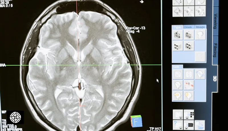
T1 / T2 / FLAIR / DWI / SWI — what each sequence sees
A standard screening brain MRI is not a single image but a stack of sequences, each tuned to different tissue properties. Understanding the basics helps you read your own report — and helps you spot whether a center is actually running a thorough protocol or a stripped-down version.
| Sequence | What it shows | Best for detecting |
|---|---|---|
| T1-weighted | Anatomic detail; fat bright, fluid dark | Structural anatomy, atrophy, post-contrast tumor enhancement |
| T2-weighted | Fluid bright, edema bright | Cysts, edema, tumors, inflammation |
| FLAIR | T2 with cerebrospinal fluid suppressed | White matter hyperintensities, MS plaques, small vessel disease |
| DWI | Restricted diffusion of water molecules | Acute ischemic stroke (within minutes to hours) |
| SWI | Magnetic susceptibility — iron, blood products | Microbleeds, cavernous malformations, calcifications |
| 3D T1 (MPRAGE) | High-resolution volumetric anatomy | Hippocampal volumetry, surgical planning, atrophy quantification |
| DIR | Double inversion recovery — suppresses both CSF and white matter | Cortical and juxtacortical MS lesions |
| TOF-MRA | Time-of-flight angiography — flowing blood | Aneurysms, AVMs, vessel stenosis (no contrast required) |
A reasonable screening brain protocol includes at minimum T1, T2, FLAIR, DWI, and SWI. If the radiologist or clinician adds 3D MPRAGE for volumetry or TOF-MRA for vascular screening, that's a more thorough exam. If a "brain MRI" reports only T2 and FLAIR, you're getting a partial picture.
3T benefit for brain specifically
Field strength matters more for brain than for almost any other organ. The signal-to-noise ratio at 3 Tesla is roughly twice that of 1.5T, and you can spend that extra signal in two ways: thinner slices (better small-lesion detection) or shorter scan times (better patient tolerance, fewer motion artifacts). For brain imaging, radiologists generally bank the gains as resolution.
The clinically meaningful differences at 3T:
- Sub-3mm lesion visibility. Aneurysms under 3mm, small cavernous malformations, and tiny demyelinating plaques are reliably visible at 3T but easily missed at 1.5T. The MAGNIMS consensus on multiple sclerosis imaging explicitly recommends 3T for surveillance because plaque counts and burden assessment are more accurate.
- Microbleed detection. Susceptibility-weighted imaging (SWI) at 3T picks up microbleeds — sub-millimeter deposits of hemosiderin from prior microscopic vessel rupture — far more sensitively than 1.5T. Microbleed burden is increasingly used in cerebral amyloid angiopathy, hypertensive small vessel disease, and CADASIL workup.
- Subclinical white matter changes. The early stippling of white matter hyperintensities that precedes clinical small vessel disease is more confidently characterized at 3T, where the contrast-to-noise advantage of FLAIR is most pronounced.
- Hippocampal volumetry. Quantifying hippocampal volume — a key biomarker in early Alzheimer's risk stratification — requires the sub-millimeter isotropic resolution that 3D MPRAGE at 3T delivers cleanly.
For a healthy adult getting a one-time baseline, the practical question is not "does 3T find more?" but "do I want the version of the scan that finds things at 3mm or the version that finds them at 8mm?" Most people, told it that way, choose 3mm.
MR Angiography — when aneurysm screening makes sense
MR Angiography (MRA) is a different question from a structural brain MRI. Standard brain MRI shows you tissue. MRA shows you the vessels. Most centers use time-of-flight (TOF) MRA, which exploits the signal from flowing blood and requires no contrast injection. It's added to a brain protocol in roughly 10 minutes.
The case for adding MRA depends almost entirely on risk profile. The general-population prevalence of unruptured intracranial aneurysms is around 3%, but the relevant subgroups carry meaningfully higher risk:
- First-degree family history of subarachnoid hemorrhage or known aneurysm. Roughly 1.4x to 4x increased risk depending on the number of affected relatives. Two or more first-degree relatives with aneurysmal SAH is a strong indication for screening.
- Autosomal dominant polycystic kidney disease (ADPKD). Approximately 10% lifetime aneurysm prevalence. Most ADPKD guidelines recommend screening MRA in carriers, particularly with a family history of rupture.
- Connective tissue syndromes — Ehlers-Danlos vascular type, Loeys-Dietz, Marfan with arterial involvement.
- Heavy smokers and patients with poorly controlled hypertension in their 40s and beyond — particularly women, who have higher aneurysm prevalence than men.
The hard part is what happens if you find one. The ISUIA (International Study of Unruptured Intracranial Aneurysms) and follow-up cohorts established the framework most clinicians still use: aneurysm size and location predict rupture risk. Anterior circulation aneurysms under 7mm in patients with no prior SAH have very low annual rupture rates (well under 1%); posterior circulation aneurysms and larger lesions carry meaningfully higher rates. The pragmatic rule of thumb most neurosurgeons follow:
- Under 7mm, anterior circulation, asymptomatic, no family history of rupture — typically observation with surveillance MRA at 6-12 months, then annually.
- 7mm and above, or any size in posterior circulation, or any size with concerning morphology (irregular, daughter sac) — refer to a neurovascular team for treatment discussion (clipping vs endovascular coiling vs flow diverter).
If you're not in a screening-indicated subgroup, adding MRA is reasonable but optional. If you are, it's the most valuable 10 extra minutes you'll spend in the magnet.
Common incidental findings — what they typically mean
Any time you image a healthy population thoroughly, you find things. The literature on incidental brain MRI findings in asymptomatic adults — most prominently the Rotterdam Scan Study — found that something flagged as "abnormal" appears in roughly 15-20% of healthy adults, but the vast majority require nothing more than awareness or a follow-up scan.
- Unruptured aneurysms (~3% prevalence). The majority are small (<7mm), anterior circulation, and managed with surveillance. Found incidentally and observed, they are usually a non-event.
- Arachnoid cysts (~1-2%). Benign CSF-filled pockets, almost always asymptomatic, requiring no treatment unless they exert mass effect — which is rare.
- Pituitary microadenomas (~10% in autopsy series). Sub-1cm pituitary lesions are common and most are non-functioning. Endocrine workup (prolactin, cortisol, IGF-1) is the standard response, and most are managed with monitoring.
- Cavernous malformations (~0.5%). Clusters of abnormal capillaries that may bleed slowly. Most asymptomatic ones are observed; symptomatic or repeatedly bleeding lesions may need surgery.
- Vestibular schwannomas (~0.02%). Benign tumors on the vestibulocochlear nerve. Small, asymptomatic ones are observed; growth or hearing changes prompt treatment discussion.
- Chiari I malformation (~1%). Cerebellar tonsils descending into the foramen magnum. Most are asymptomatic incidental findings; surgical decompression is reserved for symptomatic cases.
- Sinusitis and mucosal thickening. Extremely common, almost always clinically irrelevant on a screening MRI.
- White matter hyperintensities. Increase with age, hypertension, smoking, and small vessel disease. A few are unremarkable in older adults; a heavy burden in someone under 50 deserves a vascular risk-factor workup.
- Microbleeds. Lobar microbleeds raise the question of cerebral amyloid angiopathy; deep microbleeds suggest hypertensive small vessel disease. Pattern matters more than count.
The right way to read an incidentaloma report is with someone who can contextualize it. A clean radiology report describing "a 2mm anterior communicating artery aneurysm, no daughter sac, recommend surveillance MRA in 12 months" is reassuring news in 2026 — it would have been invisible at 1.5T five years ago, and you now have years of warning to monitor it.
Multiple sclerosis surveillance
For patients with established or suspected multiple sclerosis, brain MRI isn't optional — it's the central tool for diagnosis, monitoring, and treatment decisions. The MAGNIMS consensus (the European MS imaging working group) lays out the standard:
- 3T is now the recommended field strength for MS surveillance, with 1.5T accepted but considered inferior.
- High-resolution 3D FLAIR is the workhorse sequence for plaque detection.
- Double inversion recovery (DIR) substantially improves detection of cortical and juxtacortical lesions, which are diagnostically important and easily missed on FLAIR alone.
- Annual surveillance is standard for stable patients; more frequent imaging is used during treatment changes or new symptoms.
If you're an MS patient considering imaging in Taiwan, confirming that the partner hospital runs a MAGNIMS-aligned protocol with DIR is the right diligence question.
Cognitive aging baseline — when it's worth establishing
This is an emerging use case that the longevity community has pushed into the mainstream. The argument: hippocampal atrophy precedes clinical Alzheimer's by 10-15 years. If you have a family history of dementia, establishing a quantified baseline in your 40s or 50s gives you a comparison point that's far more informative than an absolute volume read in your 70s.
Practically, this requires a 3D MPRAGE sequence with sub-millimeter resolution (standard at 3T) and either software-based or manual hippocampal volumetry. Some Taiwan centers will run automated NeuroQuant or FreeSurfer-style analysis on the volumetric data; others provide the raw images and leave volumetry to a specialist. Ask before you book.
Is this for everyone? No. For most healthy adults without family history, the marginal value of cognitive volumetry is low — you're unlikely to act on it for decades. For someone with an APOE4 allele, two affected parents, or active cognitive concerns, a baseline is one of the more defensible investments in personal data you can make.
Practical considerations: claustrophobia, implants, contrast
The honest answers to the three questions everyone has:
Claustrophobia. The bore of a modern 3T magnet is wider than the older 1.5T tunnels people remember from the 2000s, but it's still confined. Strategies that actually work, in order of effectiveness: (1) take the offered prone-vs-supine option if available — supine is standard but some centers permit feet-first positioning for brain, which keeps the head closer to the open end; (2) eye mask plus deep breathing — removing visual cues paradoxically reduces panic; (3) request mild oral anxiolytic (a single dose of lorazepam or similar) timed for the scan — most Taiwan partner hospitals can arrange this on prior request; (4) light IV sedation as a last resort. "Open MRI" units at lower field strength exist but defeat the point of going to a 3T-equipped center.
Implants. Most modern implants are MRI-conditional, meaning safe under specified conditions. The categories that need explicit clearance: cardiac pacemakers and ICDs (many newer models are MRI-conditional, but require programming changes before scanning), cochlear implants (most newer models are conditional with magnet management), deep brain stimulators and other neurostimulators (require device-specific protocols), aneurysm clips placed before 1995 (older ferromagnetic clips are absolute contraindications). Bring documentation. Joint replacements, dental work, and most orthopedic hardware are not problematic.
Contrast. Gadolinium-based contrast agents (GBCAs) are not used in routine screening brain MRI. They're reserved for tumor characterization, infection workup, and detailed MS activity assessment. The risk profile of modern macrocyclic GBCAs is favorable, but skipping unnecessary contrast is the right call when the indication is screening rather than known disease.
Pricing comparison — Taiwan partner vs Prenuvo/Ezra/U.S. hospital
Brain MRI pricing varies more by where you buy it than by what you actually get. The same 3T scanner running the same protocol is priced very differently across U.S. hospital radiology, U.S. screening services, and Taiwan partner hospitals.
| Provider | Cash price (USD) | What's included |
|---|---|---|
| U.S. hospital radiology (dedicated brain MRI) | $1,800 - $3,200 | Brain MRI only, radiologist read, no MRA unless ordered separately |
| U.S. outpatient imaging center | $700 - $1,500 | Brain MRI only; quality varies, often 1.5T |
| Prenuvo | $2,499 (whole-body, brain included) | Whole-body screening MRI; brain coverage is basic |
| Ezra Full | $1,500 - $2,500 | Multi-organ MRI screening; brain included |
| Taiwan partner single-region MRI (no contrast) | $310 | Full multi-sequence 3T brain protocol with English report |
| Taiwan partner single-region MRI (with contrast) | $550 | Brain with contrast; TOF-MRA bundled into Advanced + Brain MRA package |
| Taiwan full-body package (brain bundled, Light tier) | $1,399 | Brain MRI as part of complete 2-hour morning protocol — see our service packages |
| Taiwan Advanced + Brain MRA package | $3,799 | Full-day workup including brain MRI + MR angiogram + comprehensive imaging |
The arbitrage is real. A standalone brain MRI plus MRA at a Taiwan partner hospital costs roughly what a co-pay would in the U.S. — and the underlying scanner, sequence library, and radiologist subspecialty training are at parity. Our overview of why Americans are flying to Taiwan for full-body MRI walks through the broader economics.
How brain MRI fits into a Taiwan screening morning
At our partner hospital in Beitou, brain imaging slots into a 4-hour morning protocol that pairs MRI with a comprehensive workup including bloods, ultrasound, and cardiology screening. The practical sequence:
- Arrive 7:30am, intake and bloods first — fasting bloods come early, before contrast questions matter.
- Ultrasound and cardiology while bloods process — abdominal, thyroid, carotid Doppler, echo as indicated.
- MRI block, 60-90 minutes total. Brain MRI itself is 25-30 minutes when bundled within a full-body protocol; if you've requested a dedicated detailed brain protocol with MPRAGE volumetry and DIR, plan on 45-60 minutes for brain alone.
- Add MRA if indicated — adds about 10 minutes, no contrast, decided ahead of the appointment based on family history and risk profile.
- Contrast is the exception, not the default — only added when there's a specific tumor or active disease question, which is rare in a screening context.
- Same-day preliminary read, full radiology report within 48 hours in English. AI-augmented review by partner radiology improves consistency on small lesions — see our piece on AI in Taiwan health screening.
If you're traveling specifically for brain imaging, a single morning gives you a multi-sequence 3T study plus the option of MRA, with a board-certified neuroradiology read and English-language report — typically for less than the cost of the U.S. radiology bill alone, before insurance and before factoring travel.
For full booking workflow, see our partner provider directory. For deeper context on the underlying scanner technology, our 3T standard in Asia article covers the hardware side.
Sources & Further Reading
- USPSTF — U.S. Preventive Services Task Force recommendations
- American Cancer Society — Cancer Facts & Figures
- NCI SEER — Surveillance, Epidemiology, and End Results Program
- CDC — Cancer prevention & screening
- American Heart Association — heart attack risk
- American College of Cardiology — clinical guidelines
FAQ
When brain imaging is bundled inside a full-body screening protocol at our Taiwan partner hospital, the brain block itself runs 25-30 minutes. A dedicated detailed brain protocol — with high-resolution 3D MPRAGE for volumetry, DIR for MS surveillance, and full multi-sequence coverage — runs 45-60 minutes. Adding MR Angiography (TOF-MRA) is an extra 10 minutes and requires no contrast.
It depends on your risk profile. Add MRA if you have a first-degree relative with subarachnoid hemorrhage or a known aneurysm, autosomal dominant polycystic kidney disease (ADPKD), a connective tissue syndrome (Ehlers-Danlos vascular type, Loeys-Dietz), or are a heavy smoker with poorly controlled hypertension. Without those risk factors, the general-population aneurysm prevalence is around 3% and routine MRA screening is optional rather than indicated.
The ISUIA framework is the standard. Anterior circulation aneurysms under 7mm in patients with no prior subarachnoid hemorrhage have very low annual rupture rates (well under 1%) and are typically managed with surveillance MRA at 6-12 months, then annually. Aneurysms 7mm and above, posterior circulation lesions, or those with concerning morphology (irregular shape, daughter sac) prompt referral to a neurovascular team to discuss clipping, endovascular coiling, or flow diversion. Most incidentally found small aneurysms are observed, not treated.
No. Gadolinium-based contrast agents (GBCAs) are reserved for tumor characterization, infection workup, and detailed MS activity assessment. For a routine screening brain MRI in a healthy adult, contrast is not used. Modern macrocyclic GBCAs have a favorable safety profile, but the right principle is to skip what you do not need — a multi-sequence non-contrast 3T brain protocol covers the screening question completely.
Yes, and it is more manageable than people expect. Modern 3T magnets have wider bores than older units. Effective strategies in order: an eye mask plus deep breathing, music or noise-cancelling headphones, a single oral anxiolytic dose (lorazepam or equivalent) timed for the scan — Taiwan partner hospitals can arrange this with prior request — and IV sedation as a last resort. "Open MRI" units exist but operate at lower field strength and defeat the purpose of choosing a 3T center.
Prenuvo bundles brain coverage inside its $2,499 whole-body screening protocol (Prenuvo pricing as of 2026-05). The brain portion is real but relatively basic — adequate for gross structural screening, less optimized for sub-3mm lesion detection, MS plaque characterization, or hippocampal volumetry. A standalone Taiwan partner single-region MRI (no contrast) runs $310 with the same multi-sequence T1/T2/FLAIR/DWI/SWI protocol at 3T; the Advanced + Brain MRA package ($3,799) adds TOF-MRA plus a full-day workup. The Prenuvo proposition is convenience and U.S. location; the Taiwan proposition is depth at a fraction of the price. Prices reflect 2026-05 — see /services for current.
For most healthy adults without family history of dementia, the marginal value is low — you are unlikely to act on the data for decades. For someone with an APOE4 allele, two affected parents, or active cognitive concerns, a quantified baseline in your 40s or 50s is one of the more defensible longevity investments. It requires a 3D MPRAGE sequence with sub-millimeter resolution (standard at 3T) and either automated volumetry software (NeuroQuant, FreeSurfer-style) or specialist manual analysis. Confirm with the center in advance whether volumetry is included in the report or requires separate processing.