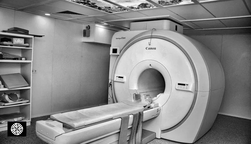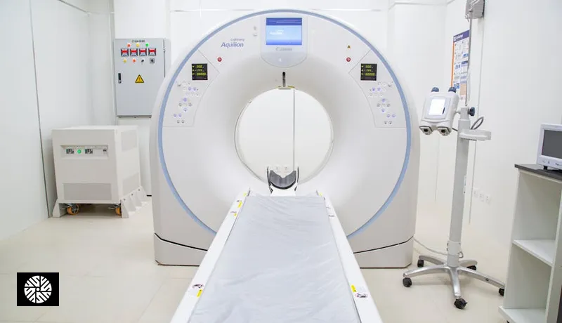

"3T" is a specification, not a diagnosis: it is the static magnetic field strength of the scanner, twice the 1.5T of the machines still in routine clinical use worldwide. The extra field strength is real and it helps with some scans more than others. It is also the easiest thing for a clinic to put on a brochure, which is why it gets quoted far more often than the two things that change a report more — the protocol that was run and the radiologist who read it. Below: what the radiology literature and the manufacturers actually claim for 3T, what they do not claim, and what an MRI costs with us.
Is 3T better than 1.5T?
For prostate MRI, the question has a documented answer. PI-RADS v2.1 — the international standard for how prostate MRI is acquired and reported — accepts both 1.5-T and 3-T systems and prefers 3-T "because of its superior signal-to-noise ratio, which can be leveraged to improve spatial and temporal resolution". Preferred, not required.
That is the honest shape of the whole 3T argument. More signal, which a good protocol spends on either finer detail or a shorter scan. And the gap is narrowing rather than widening: the same review notes that better external phased-array coils, optimised protocols and deep-learning reconstruction have closed much of the image-quality distance between the two field strengths. A 1.5T scan run on a complete protocol and read by a subspecialist beats a 3T scan that skipped sequences.
What does PI-RADS v2.1 require of the scanner?
Prostate MRI is the application where acquisition is most tightly specified, so it is a useful yardstick for what "a proper MRI" means. These are the requirements as summarised in the current review literature:
| Parameter | What PI-RADS v2.1 asks for |
|---|---|
| Field strength | 1.5-T and 3-T both acceptable; 3-T preferred for its higher signal-to-noise ratio |
| Endorectal coil | Optional — modern external coil designs make it less necessary |
| T2-weighted imaging | Axial plus one orthogonal plane; 2D mandatory, 3D an optional adjunct |
| Diffusion-weighted imaging | At least two b-values (50–100 and 800–1000 s/mm²), plus a high b-value of ≥1400 s/mm², acquired or synthesised |
| ADC map | Calculated from b-values ≤1000 s/mm² |
If you are booking a prostate MRI anywhere in the world, that table is the checklist. "We have a 3T" answers one row of it.
What do the scanner software packages actually change?
Most of the recent gain in MRI has come from reconstruction and coil software rather than from bigger magnets. Here is what the three major manufacturers claim, in their own words on their own product pages — vendor claims measured under vendor conditions, so read them as the ceiling rather than the average:
| Manufacturer | Feature | Manufacturer's own claim |
|---|---|---|
| GE HealthCare | AIR Recon DL — deep-learning image reconstruction | Improved signal-to-noise ratio "means scan times are cut by up to 50%"; "sharpens images by up to 60%" |
| Philips | Compressed SENSE — accelerated acquisition | "Up to 50% faster MRI scans with virtually equal image quality", or "up to 64% higher spatial resolution within the same scan time" |
| Siemens Healthineers | MAGNETOM Vida — 3T platform with BioMatrix sensors | 3 Tesla with a "70 cm / 2.30 ft Open Bore design"; BioMatrix adjusts the exam to patient variability |
Two practical consequences. A wide bore is not a 1.5T-only comfort feature — the Vida is a 70 cm bore at 3T. And those scan-time claims are what makes a stacked screening session possible: time saved on the easy regions is what buys room for the demanding ones in a single visit.
How much does an MRI cost at New Dawn?
Our MRI prices are listed in full on /packages. Single scans and add-ons:
| Scan | Our price (USD) |
|---|---|
| Single-region MRI, no contrast | $310 |
| Single-region MRI with contrast | $550 |
| Comprehensive Breast Imaging — breast MRI with contrast, mammogram and breast ultrasound | $619 |
| Whole Spine MRI | $699 |
| Brain MRA | $699 |
If the question is broader than one body region, the full-body packages cost less than stacking single scans: Light at $1,399 covers head, neck, abdomen, pelvis and whole spine by MRI without contrast, plus a low-dose lung CT and ultrasounds; Complete ($1,699) adds a DEXA scan and a coronary calcium score CT; Plus ($3,099) adds CT angiography, a brain MRA and a carotid ultrasound. If a brain MRA is the specific reason for the trip, the Advanced Package with Brain MRA is $3,799. Every price includes the radiologist's report and a doctor's debrief in the same visit.
For the comparison against US screening MRI services, see Prenuvo, Function and Ezra compared with Taiwan and why Americans fly to Taiwan for full-body MRI.
Which scanner will actually be used?
Ask, and expect a specific answer. We do not publish a partner hardware inventory on this page: scanners get replaced and software gets updated on the clinic's schedule, and a stale spec sheet is worse than none. What we do instead is confirm in writing, with the clinic, the make, model and field strength assigned to your booking before you pay. Hospital equipment pages rarely state this, which is exactly why it is worth asking of any provider, anywhere.
The reason to bother: a home physician who knows the exact platform and protocol can often use the images directly instead of re-scanning you. Ask for the raw study, not only the report.
When is 1.5T the right choice?
- Implanted devices. RadiologyInfo.org — published jointly by the Radiological Society of North America, the American College of Radiology and the American Society of Radiologic Technologists — states that some newer pacemakers, ICDs, neurostimulation systems, cochlear implants and medication pumps are acceptable for MRI, but that "the MRI technologist and radiologist must know the exact type that you have to follow special procedures to ensure your safety". The device's own labelling names the conditions it was tested under, field strength included. Bring the implant card, not a memory of it.
- Availability. PI-RADS v2.1 accepts 1.5-T, and so does most screening imaging. A 1.5T appointment next week on a complete protocol is worth more than a 3T appointment next quarter.
- Claustrophobia. Bore width is a separate specification from field strength — ask for the bore in centimetres rather than assuming the lower-field scanner is the roomier one.
What does field strength not fix?
The same scanner produces different reports depending on:
- Sequence selection. Skipping sequences saves time and loses information. The PI-RADS table above is what a fully specified protocol looks like; ask for the equivalent for your body region.
- Radiologist subspecialty. Ask whether the reading radiologist has a fellowship in body, neuro or breast imaging, matched to your question.
- Report structure. PI-RADS and BI-RADS scores, lesion measurements with reference points, and comparison against prior imaging belong in structured fields, not buried in narrative prose.
- Preparation and positioning. Coil placement, breath-holding compliance and bowel preparation all move image quality. The technologist matters.
- AI second reads. A second-read tool is a useful signal when it is named and regulatory-cleared. Ask which platform and which clearance; "we use AI" on its own is not an answer.
What should you ask on the booking call?
- "What make, model and field strength is the scanner?" — a specific make, model and tesla figure. "A modern MRI" or "high-field" is a yellow flag.
- "Which sequences are in the protocol for my question?" — a named list. For prostate, the PI-RADS v2.1 rows above; for other regions, an equivalent named set.
- "Who reads the scan, and what is their subspecialty?" — a named radiologist or team with a relevant fellowship.
- "Do I get the raw images, not just the report?" — you want the study itself, so your own physician can use it without repeating it.
- "Is a second read used, human or AI, and which?" — optional, but a specific answer is a positive signal.
For most preventive screening, protocol completeness and radiologist experience outrank the number on the magnet. Where field strength is most worth asking about: prostate, breast and brain imaging. To match a package to the question you actually have, the MRI options are on /packages and partner detail is on /providers.
Sources
- Cancer Imaging — The evolving PI-RADS paradigm (PI-RADS v2.1 field strength, coils and b-value requirements) (accessed 2026-09-08)
- GE HealthCare — AIR Recon DL (accessed 2026-09-08)
- Philips — Compressed SENSE (accessed 2026-09-08)
- Siemens Healthineers — MAGNETOM Vida 3T (accessed 2026-09-08)
- RadiologyInfo.org (RSNA, ACR, ASRT) — MRI Safety (accessed 2026-09-08)
US / UK terms and the closest Taiwan context
Read this first: These are comparison aids, not claims that coverage, credentials, or care pathways are equivalent. Europe is not one health system; UK examples below refer specifically to NHS England.
Radiology report, DICOM, and PACS
- What it means there
- A radiology report is the clinician’s interpretation. DICOM (Digital Imaging and Communications in Medicine) is the international standard for medical images and related information. PACS is a system used to store and retrieve imaging data.
- Closest Taiwan context
- Taiwan facilities may use the same international imaging standards, but the available export medium, viewer, report language, and delivery time are facility-specific.
- Do not treat as equivalent
- Receiving a PDF report does not necessarily mean the diagnostic images are included. Receiving DICOM files does not replace a signed clinical report or guarantee that another facility can import every file.
- What to ask
- Ask separately for the signed report, report language, complete DICOM study, delivery method, viewer needs, release timing, and any fee.
Frequently asked
Is 3T MRI strictly better than 1.5T?
For most preventive screening applications — brain, breast, prostate, small vessels — yes, 3T provides higher signal-to-noise ratio and better resolution of small structures. For some applications (large abdominal masses, motion-sensitive cardiac cine imaging, MRI-conditional implants approved only at 1.5T), 1.5T is comparable or sometimes preferred. Our partner network defaults to 3T for screening protocols, with 1.5T available where indicated.
Can I request a specific scanner model?
Yes — we list specific MRI hardware per partner hospital on the provider profile pages. If your physician has recommended a specific platform (e.g. Siemens MAGNETOM for prostate multiparametric MRI), we can route your booking to the matching deployment. Mention this on your concierge intake call.
Are these the same scanners U.S. centers use?
Yes. Siemens MAGNETOM Vida and Lumina, GE SIGNA Premier, and Philips Ingenia Elition are the same flagship 3T platforms deployed at Mayo Clinic, Cleveland Clinic, MD Anderson, Mass General, and major European centers like Hirslanden and Charité. In several cases the Taiwan partner deployment is a newer-generation install than the Western academic center it's compared with. Hardware quality is the easy part of imaging — the harder parts are protocol design and radiologist experience, both of which we vet for each partner.
What if I am claustrophobic — can I do 1.5T instead?
Yes. Several partners offer wide-bore 1.5T scanners (70cm bore or larger), which feel substantially less confining than a standard 60cm 3T bore. The Philips Ingenia Elition 3T deployment also has ambient lighting and audio designed to reduce claustrophobia. For severe claustrophobia, oral anxiolytic medication or — rarely — IV sedation is available. Tell us at the intake call and we will route to a comfort-optimized scanner and protocol.
Do all your partners have 3T scanners?
The major preventive-medicine partners listed on our /providers page have 3T scanners as their primary MRI platform. Some also have 1.5T scanners for specific use cases (claustrophobic patients, MRI-conditional implants approved only at 1.5T, certain cardiac protocols). Smaller specialty clinics in our network may deploy 1.5T as their primary system, which is entirely appropriate for many applications. We disclose specific hardware per partner.
How much does scanner generation matter — is a 2017 Vida the same as a 2023 Vida?
Generation matters more than most patients realize. A 2023-installed MAGNETOM Vida benefits from updated coil arrays, current software releases, and newer deep-learning reconstruction packages compared with a 2017 install — even at the same nominal 3T field strength. That said, a well-maintained 2017 Vida on current software is still excellent. Most of our partner deployments are post-2019 generation, several with hardware refreshes in the past three years.
Is open-bore MRI available?
True "open MRI" units (typically 0.7T or 1.0T with no bore) are not part of our preventive screening network — image quality is materially lower for the screening applications that matter most (brain, prostate, breast). However, several partners offer wide-bore 3T (60cm) and wide-bore 1.5T (70cm) scanners that are far less confining than older 55cm-bore designs. For most patients with mild-to-moderate claustrophobia, a wide-bore 3T plus a brief anxiolytic is preferable to a low-field open scanner.