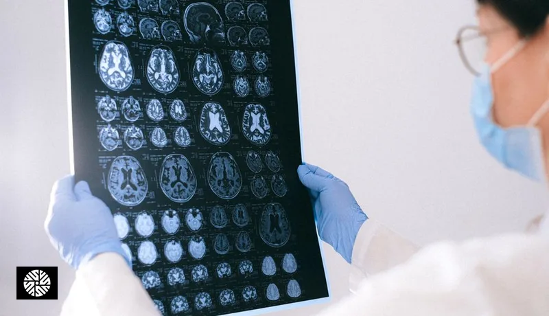

Visceral fat is the fat packed around the abdominal organs, as opposed to the fat under the skin. Its link to diabetes and heart disease is well established. A growing set of imaging studies now asks a second question: does visceral fat in midlife show up in the brain too?
Why does visceral fat get more attention than other fat?
In the Framingham Heart Study, 3,001 adults without heart disease had both kinds of abdominal fat measured by CT.
Per standard-deviation increase, visceral fat was associated with 4.7-fold odds of metabolic syndrome in women and 4.2-fold in men, against 3.0 and 2.5 for fat under the skin.
Visceral fat stayed more strongly associated with an adverse metabolic profile even after accounting for BMI and waist size.
Our visceral fat guide covers the basics of what it is and how it is measured.
Is visceral fat linked to a smaller brain?
An early large study was also from Framingham. In 733 middle-aged adults with a mean age of 60, more visceral fat on CT was associated with lower total brain volume.
The association held after adjusting for BMI and for insulin resistance.
It weakened and was no longer statistically significant after adjusting for C-reactive protein, a marker of inflammation, which suggests inflammation may be part of the pathway.
In 2024, a study of 10,001 healthy adults with a mean age of 52.9 used brain and abdominal MRI read by deep-learning software.
Visceral fat correlated with lower grey matter volume (r = −0.44), lower white matter volume (r = −0.41) and a smaller hippocampus (r = −0.39).
Fat under the skin was also related to lower brain volumes, so abdominal fat in general, not only visceral fat, tracked with the result.
Both studies are cross-sectional: they show that the two travel together, not that fat shrinks the brain.
Does midlife belly fat raise dementia risk?
In a Kaiser Permanente cohort, 6,583 members had their sagittal abdominal diameter (belly depth, front to back) measured between 1964 and 1973, and 1,049 of them were diagnosed with dementia about 36 years later.
Those with the largest abdominal measurements had a 2.72-fold hazard of dementia compared with the smallest, and a 1.92-fold hazard after accounting for BMI.
People who were both obese and had a large abdomen had a 3.60-fold hazard.
The authors concluded that central obesity in midlife increases dementia risk independent of diabetes and cardiovascular disease.
A small 2024 pilot study of 32 cognitively normal adults in midlife found that a higher ratio of visceral to subcutaneous fat went with more amyloid, the protein linked to Alzheimer's disease, in one brain region; the link was significant in men but not in women.
| Study | People | How fat was measured | Main finding |
|---|---|---|---|
| Framingham, 2010 | 733, mean age 60 | CT | More visceral fat, lower total brain volume |
| Raji et al., 2024 | 10,001, mean age 52.9 | MRI with deep learning | Visceral fat vs grey matter r = −0.44 |
| Kaiser Permanente, 2008 | 6,583, followed about 36 years | Sagittal abdominal diameter | Largest vs smallest: dementia hazard 2.72 |
| Dolatshahi et al., 2024 (pilot) | 32, midlife | MRI | Higher visceral-to-subcutaneous ratio, more amyloid in men |
How is visceral fat measured outside a research study?
CT and MRI measure it directly, but a DEXA body-composition scan estimates it with much less effort.
In 124 adults aged 18 to 90, DEXA visceral fat agreed closely with CT, with an r² of 0.957.
Waist size is the free first check: the NHLBI says a waist over 35 inches for women or 40 inches for men increases risk, which is about 89 cm and 102 cm.
In the same DEXA study, BMI and waist size were only poor to modest guides to the visceral fat number itself.
Can visceral fat be reduced?
Yes. A meta-analysis of 117 studies and 4,815 participants found that both exercise and calorie-reduced diets reduce visceral fat.
Diet produced more weight loss, while exercise tended to cut more visceral fat.
Without weight loss, exercise was associated with a 6.1% fall in visceral fat, against 1.1% for diet.
Scale weight alone can therefore miss real progress; the authors called total weight loss a poor marker of visceral fat change.
Whether reducing visceral fat in midlife protects the brain has not been tested in a trial; the evidence above is observational.
Where does visceral fat fit in a screening visit?
The DEXA Total Body & Bone Health Package ($109) gives a visceral fat estimate along with body composition and bone density.
Our Complete package ($1,699) includes that DEXA scan with a full-body MRI.
For brain imaging options and what they can and cannot show, see our guide to normal brain aging.
The full range is on our health screening page.
Sources
- Fox et al. — Abdominal visceral and subcutaneous adipose tissue compartments: association with metabolic risk factors in the Framingham Heart Study, Circulation 2007 (accessed 2026-10-29)
- Debette et al. — Visceral fat is associated with lower brain volume in healthy middle-aged adults, Annals of Neurology 2010 (accessed 2026-10-29)
- Raji et al. — Deep learning-derived abdominal fat and brain volumes in 10,001 adults, Aging and Disease 2024 (accessed 2026-10-29)
- Whitmer et al. — Central obesity and increased risk of dementia more than three decades later, Neurology 2008 (accessed 2026-10-29)
- Dolatshahi et al. — Alzheimer Disease Pathology and Neurodegeneration in Midlife Obesity: A Pilot Study, Aging and Disease 2024 (accessed 2026-10-29)
- Kaul et al. — Dual-energy X-ray absorptiometry for quantification of visceral fat, Obesity 2012 (accessed 2026-10-29)
- NHLBI — Assessing Your Weight and Health Risk (accessed 2026-10-29)
- Verheggen et al. — A systematic review and meta-analysis on the effects of exercise training versus hypocaloric diet, Obesity Reviews 2016 (accessed 2026-10-29)
US / UK terms and the closest Taiwan context
Read this first: These are comparison aids, not claims that coverage, credentials, or care pathways are equivalent. Europe is not one health system; UK examples below refer specifically to NHS England.
Radiology report, DICOM, and PACS
- What it means there
- A radiology report is the clinician’s interpretation. DICOM (Digital Imaging and Communications in Medicine) is the international standard for medical images and related information. PACS is a system used to store and retrieve imaging data.
- Closest Taiwan context
- Taiwan facilities may use the same international imaging standards, but the available export medium, viewer, report language, and delivery time are facility-specific.
- Do not treat as equivalent
- Receiving a PDF report does not necessarily mean the diagnostic images are included. Receiving DICOM files does not replace a signed clinical report or guarantee that another facility can import every file.
- What to ask
- Ask separately for the signed report, report language, complete DICOM study, delivery method, viewer needs, release timing, and any fee.