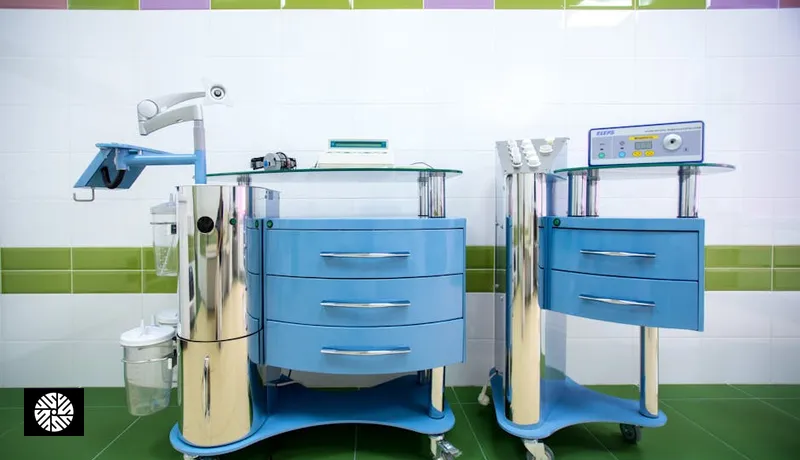
Most other regions on this site are read by a machine from the outside. Here a camera goes in, because the lining is where the trouble starts.
What gets checked, and with what
| Organ | What the test looks for | The test that does it |
|---|---|---|
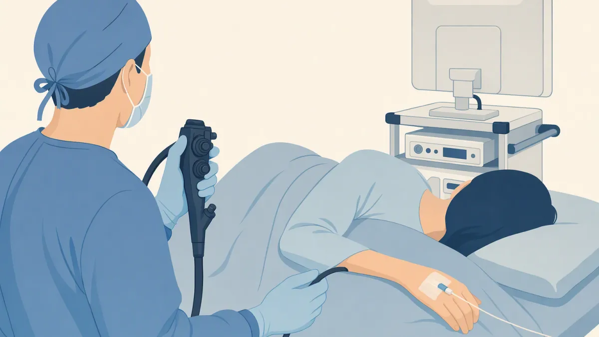
| Esophagus | Inflammation, Barrett's changes (reflux-damaged lining that can turn precancerous) and early cancer | Gastroscopy — a thin camera passed down from the mouth |
| Stomach | Gastric cancer, and Helicobacter pylori — a common, treatable infection that raises that risk | The same gastroscopy, which can take biopsies; or a stool test for H. pylori |
| Colon | Precancerous polyps and colorectal cancer | A stool test (FIT), or a colonoscopy, which can remove many polyps it finds |
Esophagus
- Why it matters: long-standing reflux is what drives the Barrett's changes worth catching early, so persistent heartburn is the usual reason to look closely.
- Imaging can't: read the surface. A full-body MRI or a CT shows how thick the wall is, not the lining where reflux damage and early tumors begin.
Stomach
- In Taiwan: there is no population gastroscopy program. From 2026 the government funds a one-time H. pylori stool test at 45–74 instead, because the infection raises stomach-cancer risk and can be treated.
- Elsewhere in East Asia: Korea's national program offers gastric screening every two years from 40, and Japan's guidelines from 50.
- Same scope: the camera that covers the esophagus continues into the stomach. Biopsies for H. pylori can be taken in the same procedure; the result comes from the lab later.
Colon
- Why it matters: most colorectal cancers start as polyps, but only some polyps ever become cancer. Removing them during colonoscopy lowers the risk; it does not remove it.
- Stool test first: in Taiwan, colon screening starts with a FIT, which looks for hidden blood. FIT cannot remove anything, and a positive result needs a colonoscopy.
- Even a good exam misses some: in tandem studies (two colonoscopies done back to back), about a quarter of precancerous polyps (adenomas) were missed on one pass (Zhao 2019). Symptoms after a normal colonoscopy still need a doctor.
- Imaging can't: match this. A scan may flag a mass once it is large; only colonoscopy inspects the full lining and takes tissue.
What this region's screening does not cover
What endoscopy gives you
- The one look no scanner can give — the lining itself, at the resolution of a camera close against it
- Biopsy and polyp removal in the same procedure, in most cases
- Both scopes in one session, if your booking includes both — confirmed when you book
What it does not
- Incidental findings turn up here too — a small benign polyp, mild gastritis, a hiatal hernia noted in passing. Most are recorded and rechecked at the next interval rather than treated.
- Every polyp. Even a well-prepared colon hides some, which is why the gastroenterologist sets your next interval from the pathology.
- The solid organs the tract runs past. Liver, pancreas, gallbladder and kidneys cannot be seen from inside the bowel — see the upper abdomen.
- The middle of the small intestine, beyond the reach of both scopes. A capsule camera, swallowed like a pill, is used when a doctor needs to see it.