Blog
Blog
Heart and Lung Screening in Taiwan: Preventing the Top Causes of Death
March 14, 2026
Two diseases dominate the global mortality table: cardiovascular disease and lung cancer. Together they account for the majority of preventable adult deaths worldwide — and both respond dramatically to early detection. The technology to find them years before symptoms appear is mature, validated, and widely available. The question for most travelers and expats is not whether to look, but how to do it efficiently and at a price that doesn't punish prudence.
Taiwan has quietly become one of the most cost-effective places in the world to combine a coronary artery calcium (CAC) score, a CT coronary angiogram (CCTA) when warranted, and a low-dose lung CT — all in a single morning, on the same scanner platform, with results read by both subspecialty radiologists and AI second-readers. This guide explains what each test actually does, how to read your numbers, and how a Taiwanese cardiac-and-lung workup compares with the U.S. or EU equivalent.
Cardiovascular and lung — the two preventable killers
The World Health Organization estimates roughly 17.9 million cardiovascular deaths each year — the single largest cause of death globally and the leading cause in nearly every developed country. The American Cancer Society places lung cancer at approximately 125,000 U.S. deaths annually, more than colorectal, breast, and prostate cancer combined. Both conditions share a brutal pattern: long asymptomatic windows, then sudden clinical presentation that is often advanced or fatal.
What makes them different from many other killers is how thoroughly imaging-detectable the early phase is. Coronary atherosclerosis deposits calcium that a CT scanner reads in less than 60 seconds. Pulmonary nodules appear on a low-dose chest CT well before they would ever produce a cough or shortness of breath. The biology is on our side; the bottleneck is access and willingness.
For a broader picture of how Taiwan's preventive ecosystem compares with American practice, see Preventive Health Gaps in the US: How Taiwan Fills the Void.
Coronary calcium score — the single most reassuring test in cardiology
The coronary artery calcium (CAC) score is a non-contrast, ECG-gated CT scan of the heart. It quantifies calcified plaque in the coronary arteries using the Agatston unit, a method first published in 1990 that has aged remarkably well. The scan takes about 10 seconds of breath-hold, exposes you to roughly 1 mSv of radiation (less than a year of natural background), and produces a number that powerfully predicts your 10-year risk of a cardiac event.
The score interpretation is straightforward:
| Agatston score | Risk category | Typical action |
|---|---|---|
| 0 | Very low — "warranty period" 5-10 years | Lifestyle, no statin needed in most cases |
| 1-99 | Mild plaque burden | Risk-factor modification, statin discussion |
| 100-399 | Moderate — substantially elevated risk | Statin recommended, BP and ApoB targets tightened |
| 400+ | High — equivalent to known coronary disease | Aggressive medical therapy, consider CCTA or stress imaging |
The MESA risk equation (from the Multi-Ethnic Study of Atherosclerosis) lets clinicians plug your CAC score directly into a recalibrated 10-year ASCVD risk estimate, often reclassifying patients up or down by a meaningful margin compared with traditional Framingham-style calculators. A 55-year-old with borderline cholesterol and a CAC of 0 is genuinely low-risk; the same patient with a CAC of 300 sits in a different universe.
The 2018 ACC/AHA cholesterol guideline endorsed CAC as a tiebreaker for borderline (5-7.5%) and intermediate (7.5-20%) 10-year risk patients deciding whether to start a statin. The U.S. Preventive Services Task Force still grades the test "I" (insufficient evidence) — a position increasingly out of step with the cardiology community, largely because USPSTF demands randomized outcome trials that are arguably unethical to run when the test is already in widespread guideline use.
When CCTA adds to CAC — anatomic and functional detail
A calcium score tells you how much calcified plaque exists, but not where the lumen is narrowed or whether soft, non-calcified plaque is present. CT coronary angiography (CCTA) uses iodinated contrast and high-resolution gated CT to map the coronary tree itself — every vessel, every stenosis, every plaque morphology. It is the closest thing to a non-invasive cath lab available today.
CCTA is most useful when:
- The CAC score is intermediate or high and a clinician needs to know if any vessel has flow-limiting disease
- The patient has atypical chest symptoms but a normal stress test (CCTA has higher negative predictive value)
- There is a strong family history of premature coronary disease and a CAC of 0 — soft plaque can exist without calcification, particularly in younger patients
The newest layer is FFR-CT (fractional flow reserve from CT), commercially deployed by HeartFlow. It applies computational fluid dynamics to a CCTA dataset and produces a non-invasive estimate of whether a given stenosis is functionally significant — exactly the question that previously required a catheter and a pressure wire. Several Taiwan partners now route ambiguous CCTA findings through HeartFlow before deciding whether to proceed to invasive angiography.
Low-dose lung CT — beyond the heavy-smoker indication
The National Lung Screening Trial (NLST), published in the New England Journal of Medicine, demonstrated a 20% relative reduction in lung cancer mortality among heavy smokers screened with low-dose CT compared with chest X-ray. On the strength of NLST and the European NELSON trial, the USPSTF now grades low-dose CT a Grade B recommendation for adults aged 50-80 with a 20+ pack-year smoking history who currently smoke or quit within the past 15 years.
That eligibility framework was built on Western, predominantly smoker datasets. It badly understates the situation in East Asia.
Taiwan and the broader East Asian region show a strikingly different lung cancer epidemiology: 50-70% of lung cancer patients in Taiwan have never smoked, and the dominant histology is EGFR-mutated adenocarcinoma. Risk factors that matter more than smoking in this population include indoor radon exposure, cooking fume inhalation, second-hand smoke, ambient particulate pollution, and a strong genetic component (first-degree family history nearly doubles risk). The TALENT study (TAiwan Lung cancer screening for Never smokErs) screened 12,000+ never-smokers in Taiwan with low-dose CT and detected lung cancer at rates comparable to Western smoker cohorts — most at stage 0 or I, which is curable.
| Eligibility framework | Who qualifies | Notes |
|---|---|---|
| USPSTF (US, 2021) | Age 50-80, 20+ pack-years, current smoker or quit within 15 years | Grade B; covered by Medicare and most US insurers |
| Taiwan HPA LDCT program | Age 50-74 heavy smokers OR 45-74 with first-degree family history of lung cancer | First population-level program globally to fund never-smoker screening |
| Self-pay screening (Asian context) | Adults 40+ with any of: family history, radon exposure, chronic exposure to cooking fumes, environmental risk | Justified by TALENT trial data and East Asian EGFR epidemiology |
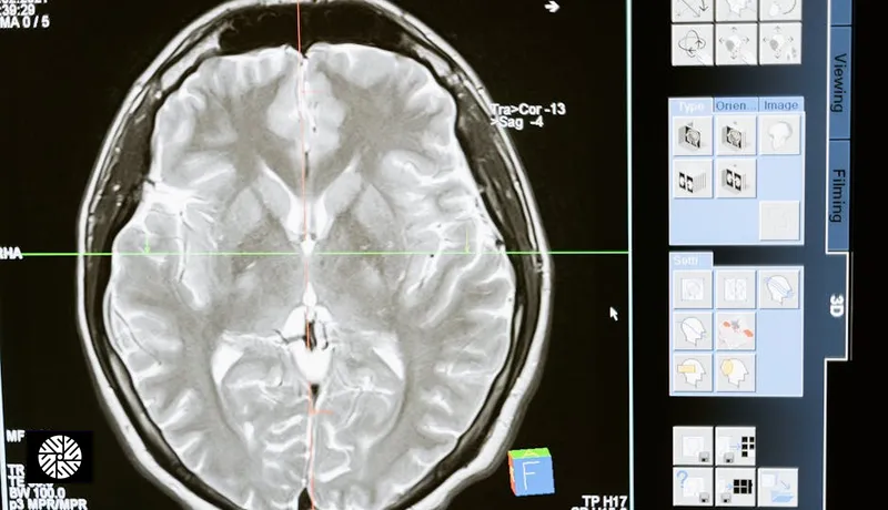
The scan itself is non-contrast, takes one breath-hold, and uses a low-dose protocol delivering roughly 1.0-1.5 mSv — comparable to six months of natural background radiation, and a small fraction of a standard chest CT. Findings are reported using Lung-RADS (1 through 4X), a structured classification that defines follow-up intervals from a routine annual scan to short-interval follow-up to tissue diagnosis. Taiwan's full-body imaging culture, including the full-body MRI workflow described in our step-by-step guide, slots low-dose lung CT in naturally as a complementary, radiation-aware add-on.
How a Taiwan workup integrates these
The practical advantage of doing this in Taiwan is sequencing. A typical morning at a partner facility looks like this:
- 07:30 — Arrival, fasting bloods drawn (lipid panel, ApoB, Lp(a), hs-CRP, basic chemistry, CEA)
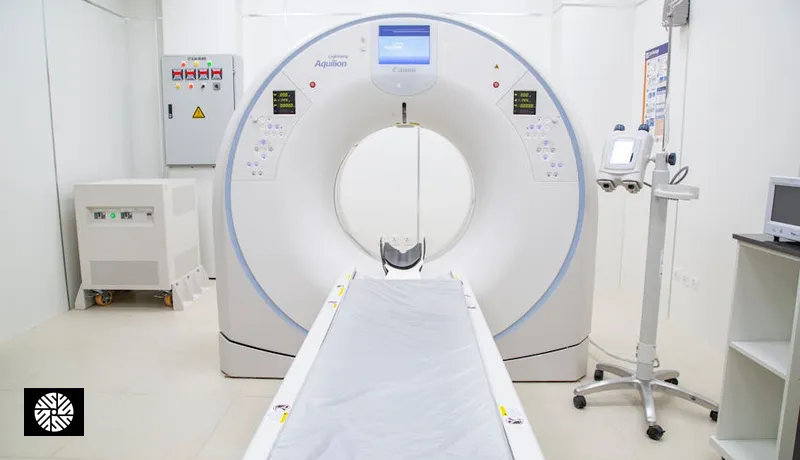
- 08:00 — CAC scan on a 320-slice CT (10 seconds of acquisition; one breath-hold)
- 08:15 — Low-dose lung CT on the same scanner (one additional breath-hold; reused gating data)
- 08:30 — If CAC is intermediate or risk factors warrant, CCTA is performed with iodinated contrast in the same sitting
- 09:00 — Resting echocardiogram if indicated by family history or athletic baseline
- 10:00 — Breakfast and physician consultation; preliminary CAC and lung CT findings often available the same morning, full reads with Lung-RADS classification within 24-48 hours
Beitou Health Management Hospital, one of New Dawn's cardiac imaging partners, runs a Siemens dual-source 320-slice scanner for coronary studies and a GE SIGNA Premier with HeartFlow integration for advanced functional cardiac imaging. The combination handles both high heart rates (no beta-blocker preparation needed for many patients) and the complex post-processing required for FFR-CT.
AI second-read on cardiac and lung CT — real-world deployment
Taiwan's hospitals were early adopters of AI-augmented radiology, partly because the country's strong domestic medical AI sector (Taipei-based companies like aetherAI, plus regional players) creates close hospital-vendor feedback loops. For a deeper look at the accuracy gains, see How AI Health Screening in Taiwan Redefines Accuracy in Asia.
For lung CT, the dominant tools are Lunit INSIGHT CXR/CT, Riverain ClearRead CT (vessel-suppression algorithm that improves nodule conspicuity), and AIDOC for incidental findings flagging. These act as a structured second reader: the radiologist reads the case first, then reviews the AI overlay, which has been shown in published validation studies to reduce missed sub-centimeter nodules by 15-30%.
For cardiac CT, HeartFlow's FFR-CT is the most clinically impactful AI integration — not a "second reader" exactly, but a derived dataset that converts an anatomic scan into a functional assessment. Several Taiwan partners route any CCTA with intermediate (40-70%) stenosis through HeartFlow before deciding whether to escalate to invasive angiography.
Biomarkers that complement imaging
Imaging tells you what is structurally present; blood biomarkers tell you what is biologically active. The pairing is more informative than either alone.
- ApoB — the single best lipid measure of atherogenic particle count. Increasingly preferred over LDL-C for risk stratification, and a target of modern lipid guidelines.
- Lp(a) — genetically determined, measured once in a lifetime. Elevated Lp(a) (above ~50 mg/dL or ~125 nmol/L) is an independent risk factor not captured by LDL or CAC, and it changes the conversation about aggressive prevention.
- hs-CRP — high-sensitivity C-reactive protein, a marker of vascular inflammation. Most useful when CAC is borderline; an elevated value reinforces the case for statin therapy.
- D-dimer — used in workup of suspected pulmonary embolism, not for routine screening.
- CEA — carcinoembryonic antigen, a non-specific tumor marker that is most useful for monitoring established lung adenocarcinoma rather than screening, but commonly included in Taiwanese health panels.
Echocardiography — when it is genuinely valuable
A resting transthoracic echocardiogram is not a routine preventive test for the average asymptomatic adult — but there are specific situations where it earns its place in a cardiac workup:
- Family history of hypertrophic cardiomyopathy (HCM) or unexplained sudden cardiac death in a first-degree relative — echo establishes baseline wall thickness and outflow tract anatomy.
- Athletic baseline — endurance athletes benefit from a baseline echo to distinguish physiologic remodeling from pathologic hypertrophy in future evaluations.
- Valvular disease screening in patients with murmurs, prior rheumatic fever, or known bicuspid aortic valve in the family.
- Pre-participation screening for patients planning intensive new exercise programs in middle age.
Patient personas — what each person learns
Persona 1: 50-year-old man with family history of MI. Father had a myocardial infarction at 56. LDL is borderline, blood pressure is well-controlled, BMI is normal. The conversation about whether to start a statin has gone in circles for years. A CAC scan returns a score of 185 — moderate plaque burden, putting him in a risk band that clearly justifies statin therapy and tighter ApoB targets. The same morning, his low-dose lung CT is clean (Lung-RADS 1). He leaves with an evidence-based reason to act on cardiovascular risk and a five-year baseline for the lungs.
Persona 2: 60-year-old never-smoker Asian female. Her mother had non-smoking lung adenocarcinoma diagnosed at stage III. She has no Western-guideline indication for lung CT — she has never smoked a cigarette in her life. Under Taiwanese epidemiology and TALENT trial logic, she is unambiguously a screening candidate. Her low-dose CT identifies a 7 mm part-solid nodule (Lung-RADS 4A), referred for short-interval follow-up. Her CAC score is 0, providing strong reassurance that her cardiovascular risk is low and statin therapy is not indicated despite a borderline cholesterol panel.
Persona 3: 45-year-old male endurance athlete. Marathon training, no symptoms, "feels great." Concerned about reports of coronary disease in apparently healthy athletes. CAC scan returns 0 — strong reassurance. A baseline echo shows mild physiologic LV hypertrophy consistent with athletic heart, documented for future comparison. Lung CT clean. Lp(a) measurement reveals a moderate elevation he didn't know about, prompting a conversation about long-term risk and aspirin discussion in a decade or two.
Pricing — Taiwan vs U.S./EU equivalent
Cost differences are not marginal. The Taiwanese self-pay market for cardiac and lung CT is among the most competitive in the developed world, partly because the technology is mature and partly because the country's high-volume preventive culture (driven by NHI co-payment dynamics for screening) keeps unit costs low.
| Test | Taiwan self-pay (USD) | U.S. self-pay (USD) | EU private (USD) |
|---|---|---|---|
| Coronary artery calcium (CAC) | $300-1,000 | $100-500 (where promo) up to $1,500 | $400-900 |
| CT coronary angiography (CCTA) | $1,500-4,000 | $3,000-8,000 | $2,000-5,000 |
| Low-dose lung CT | $300-800 | $300-1,200 | $400-900 |
| Bundle (CAC + lung CT + bloods) | $800-1,800 | $2,500-5,000+ | $1,500-3,500 |
Taiwan's bundled pricing is not just cheaper — it is structurally different. A U.S. patient typically pays for each component separately, books across multiple visits, and navigates insurance friction. In Taiwan, the screening center sells the whole morning as one product. For more on the imaging hardware that supports this, see Taiwan's 3T MRI Technology Sets the Standard in Asia.
Booking and what to expect next
New Dawn Health coordinates cardiac and lung screening packages with English-speaking partners across Taipei. Browse current options on our services page, review the imaging partners on our providers list, and reach out for a tailored sequencing recommendation based on your age, family history, and risk profile. Reports are delivered in bilingual format with physician follow-up included; if findings warrant further workup, the same partner network handles the next step on the same trip.
Sources & Further Reading
- USPSTF — U.S. Preventive Services Task Force recommendations
- American Cancer Society — Cancer Facts & Figures
- NCI SEER — Surveillance, Epidemiology, and End Results Program
- CDC — Cancer prevention & screening
- American Heart Association — heart attack risk
- American College of Cardiology — clinical guidelines
FAQ
If you are of East Asian descent, the answer is more often yes than Western guidelines suggest. The TALENT trial in Taiwan screened 12,000+ never-smokers and detected lung cancer at rates comparable to Western smoker cohorts, with 50-70% of Taiwanese lung cancer patients having never smoked. Risk factors that matter in this population include first-degree family history, radon and cooking-fume exposure, and ambient pollution. A single baseline scan in your 40s or 50s is reasonable if any of those apply.
A score above 100 means moderate plaque burden and a score above 400 is functionally equivalent to known coronary disease. The standard response is aggressive medical therapy — high-intensity statin, tighter blood pressure target, ApoB and Lp(a) measurement, lifestyle modification — and consideration of CT coronary angiography (CCTA) to map any flow-limiting stenoses. A high score is not an emergency; it is a clear signal to act. Many patients with scores of 400+ live decades on appropriate therapy.
Modern CCTA on a 320-slice or dual-source scanner with prospective ECG gating typically delivers 3-5 mSv — about a year of background radiation, similar to a transcontinental flight crew member's annual occupational dose. Coronary calcium scoring is even lower at roughly 1 mSv, and low-dose lung CT is 1.0-1.5 mSv. The cumulative dose for a complete cardiac and lung morning is well within the range that mainstream radiology considers low-risk for adult preventive imaging.
The dominant histology in East Asian populations is EGFR-mutated lung adenocarcinoma, which behaves differently from the squamous-cell, smoking-driven cancers that built U.S. screening guidelines. EGFR mutations are present in 50-60% of Taiwanese lung adenocarcinoma cases versus 10-15% in Western populations, occur disproportionately in never-smokers and women, and are highly responsive to targeted therapy when caught early. The implication is that screening criteria built on heavy-smoker datasets systematically under-include the population that most benefits in Asia.
For most asymptomatic adults, no — a resting echo is not a high-yield routine preventive test. The exceptions are meaningful: a family history of hypertrophic cardiomyopathy or unexplained sudden cardiac death, an athletic baseline you want documented before future evaluations, a known bicuspid aortic valve in the family, or a heart murmur on exam. In those situations, an echo earns its place. Otherwise, CAC and lipid biomarkers are higher-yield first steps.
A typical Taiwan partner facility runs fasting bloods at arrival, then sequences CAC, low-dose lung CT, and (if indicated) CCTA on the same scanner over roughly 60-90 minutes. Echo is added if family history warrants. Breakfast and a physician consultation follow, with preliminary findings usually available the same morning and full reads — including Lung-RADS classification and CAC percentile — within 24-48 hours. The whole sequence is designed to fit between hotel checkout and an afternoon of sightseeing.
A bundled CAC plus low-dose lung CT plus advanced lipid panel typically runs $800-1,800 in Taiwan versus $2,500-5,000+ self-pay in the U.S. and $1,500-3,500 in EU private clinics. CCTA alone is $1,500-4,000 in Taiwan versus $3,000-8,000 in the U.S. The gap is largest at the bundled, multi-test level — Taiwan sells the whole morning as one product, while U.S. patients typically pay per component across multiple visits.