Blog
Blog
Radiation-Free Full-Body MRI in Taiwan – Technology and Safety Explained
March 29, 2026
Radiation safety is the most under-discussed topic in screening medicine — and the most misunderstood. Some patients refuse all imaging because they've heard "radiation causes cancer." Others accept any scan their doctor orders without asking what the dose actually is. Both responses miss the truth, which is more nuanced and more interesting. This guide walks through the real numbers: what radiation a full-body MRI delivers (zero), what a low-dose CT actually exposes you to in millisieverts, how that compares to background radiation you absorb just by living, and what cumulative dose looks like for someone who screens annually for a decade. We'll also cover the pre-scan safety reviews — implants, pregnancy, contrast — that determine whether MRI is genuinely safe for you. See our screening services and the providers we work with for radiologist-led programs.
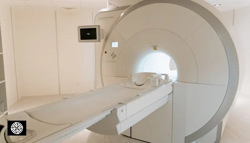
MRI is genuinely radiation-free — what that means
This is the single most important fact about MRI: it uses no ionizing radiation. Zero. None. The image is generated by a powerful magnetic field (typically 1.5 Tesla or 3 Tesla in modern scanners) and pulses of radiofrequency (RF) energy — the same type of non-ionizing energy your phone and Wi-Fi router emit, just much more focused. The hydrogen protons in your body's water molecules align with the magnetic field, get knocked off-axis by the RF pulse, and emit a tiny signal as they relax back. The scanner reads those signals and reconstructs a 3D image.
What ionizing radiation does — and MRI does not do — is knock electrons off atoms, creating ions that can damage DNA. That DNA damage is the mechanism behind radiation-induced cancer risk from X-rays, CT scans, and nuclear medicine studies. Magnetic fields and RF energy do not have enough energy to break chemical bonds. There is no mechanism by which an MRI causes cancer. Decades of large-population studies have found no link.
This is why MRI is the screening modality of choice for repeat use. You can have one every year for the rest of your life and the cumulative radiation dose remains zero. For comprehensive context on what a full-body MRI actually scans and how the workflow runs, see our full-body MRI step-by-step guide.
That said, "radiation-free" doesn't mean "consequence-free." MRI has its own safety considerations — implants, claustrophobia, contrast — which we cover below. But the cancer-from-radiation worry that makes patients hesitate before a CT? It simply does not apply to MRI.
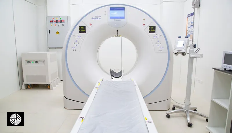
CT does have radiation — the real numbers in mSv
CT (computed tomography) uses rotating X-ray beams to build cross-sectional images. It's fast, excellent for lung nodules, calcium scoring, and emergencies — but it does deliver ionizing radiation. The dose varies enormously by scan type and protocol, and most patients have no idea what they're receiving.
The unit of measure is the millisievert (mSv), which accounts for both the energy delivered and the biological sensitivity of the tissue. Here are the typical effective doses for common CT exams:
| Scan type | Typical effective dose (mSv) | Notes |
|---|---|---|
| Full-body MRI (any field strength) | 0 | No ionizing radiation |
| Chest X-ray (single view) | ~0.1 | Equivalent to 10 days of background |
| Low-dose lung CT (NLST protocol) | ~1.5 | Screening for lung cancer in heavy smokers |
| Coronary calcium score (low-dose) | 1-3 | No contrast, gated |
| Coronary CTA (with contrast) | 5-15 | Highly protocol-dependent |
| Standard chest CT | 7-8 | Diagnostic, not screening |
| Abdominal CT | ~10 | Higher with multiphase contrast |
| PET-CT (whole body) | 15-25 | Combined CT + radiotracer |
Three things jump out. First, the difference between low-dose and standard CT is roughly 5x — protocol matters enormously. Second, modern screening protocols (low-dose lung CT, calcium score) deliver a fraction of what a diagnostic CT delivers. Third, MRI sits at zero, full stop. For more on the imaging technology side, our 3T MRI technology guide covers what differentiates Taiwan's modern equipment from older 1.5T machines.
Background radiation context — what 1 mSv actually represents
Numbers like "1.5 mSv" mean nothing without context. Here's the context: you are absorbing radiation right now, every minute of every day, from cosmic rays, soil, building materials, food, and the trace radon in the air. The global average annual background dose is around 2.4 mSv. The US average is closer to 3 mSv (higher because of indoor radon). Living in Denver, Colorado adds about 50% to your cosmic-ray dose because of altitude.
| Source | Approximate dose |
|---|---|
| Annual natural background (US average) | ~3 mSv/year |
| Annual background, high-altitude city (Denver) | ~4-5 mSv/year |
| Cross-country flight (NYC to LA) | ~0.03-0.05 mSv |
| Frequent flyer at cruise altitude | ~0.05 mSv per hour aloft |
| Living in a granite-heavy region | +0.5-1 mSv/year |
| Eating bananas (potassium-40, lifetime trivial) | ~0.0001 mSv each |
So when someone tells you a low-dose lung CT is 1.5 mSv, that's roughly six months of normal background. A calcium score at 2 mSv is about eight months. A full-body MRI is zero — you've absorbed more background just sitting in the waiting room.
Lifetime cancer risk from medical imaging — honest framing
The honest answer is that for a single low-to-moderate dose CT, the additional lifetime cancer risk is small but not zero. Population-level estimates suggest somewhere around 1 in 1,000 to 1 in 2,000 additional cancers per scan, depending on age (younger = more risk), dose, and which organs are exposed. In percentage terms, that's roughly 0.05-0.1% per scan. Compare that to the baseline lifetime cancer risk of around 40% in the general population — a single CT shifts the needle very slightly.
The framing matters in two directions. Yes, that risk is real and should not be dismissed, especially for younger patients who have more years for a radiation-induced malignancy to develop. But it should also be put alongside the diagnostic value. Refusing a clinically indicated CT to avoid a 0.05% theoretical risk while ignoring a present condition that has a 5% mortality if missed is bad math.
For screening specifically, the calculus changes again. Screening tests need to demonstrate that the population benefit outweighs both the radiation risk and the false-positive workup risk. Some pass that bar (low-dose lung CT in heavy smokers); others don't (whole-body CT in asymptomatic average-risk patients).
Low-dose lung CT specifically — NLST 20% mortality reduction outweighs the dose
The National Lung Screening Trial (NLST), published in 2011, randomized over 53,000 high-risk smokers to annual low-dose CT or chest X-ray for three years. The CT arm had a 20% reduction in lung cancer mortality and a 6.7% reduction in all-cause mortality. That's one of the largest screening benefits ever documented for any cancer.
The radiation dose per scan was approximately 1.5 mSv — comparable to six months of background. Even over a lifetime of annual screening (say, age 55 to 75, twenty scans), the cumulative dose is around 30 mSv. The estimated lifetime cancer risk from that dose is on the order of 0.1-0.3%. The screening benefit is roughly 20% relative reduction in lung cancer mortality in the eligible population, which translates to a much larger absolute benefit than the radiation harm.
This is the cleanest case in screening medicine where the math clearly favors the scan, and it's why USPSTF and other guidelines recommend annual low-dose CT for eligible smokers. For a broader look at heart and lung screening logic, see our heart and lung screening guide.
Cumulative dose for repeat screeners — annual cycle math
If you're a committed annual screener — the kind of person who books a full-body MRI plus a calcium score plus a low-dose lung CT every year — what does cumulative dose look like over a decade?
A reasonable annual stack might be:
- Full-body MRI: 0 mSv
- Brain MRI (often included): 0 mSv
- Coronary calcium score: ~1.5 mSv
- Low-dose lung CT: ~1.5 mSv
- (Optional) coronary CTA in a single year: 5-10 mSv when indicated
Total in a typical year: about 3 mSv from imaging — roughly equivalent to one year of natural background. Over ten years of annual screening, that's about 30 mSv, plus any one-off CTAs. That's meaningful but not extreme. For perspective, a single CT abdomen/pelvis with multiphase contrast can deliver 15-25 mSv in one go.
Two strategies reduce this further. First, alternate calcium score and CTA cadence — a calcium score doesn't need to be annual once you have a baseline; every 3-5 years is often enough. Second, lean on MRI wherever it can substitute. Cardiac MRI for function, abdominal MRI for liver and pancreas, prostate MRI for prostate cancer screening — all zero radiation. The annual full-body MRI carries the diagnostic load that would otherwise require multiple CTs. Our AI health screening coverage explores how AI-augmented MRI is shifting more of the workload toward radiation-free modalities.
Pregnancy and screening — what's safe
Pregnancy is the situation where radiation safety becomes acute. The developing fetus is most sensitive to radiation in the first trimester, when organogenesis is happening. Standard guidance:
- MRI without contrast: Generally considered safe in pregnancy, including the first trimester, when clinically necessary. There is no documented harm to the fetus from the magnetic field or RF energy at clinical doses. ACR guidelines support its use when needed.
- MRI with gadolinium contrast: Avoided when possible, especially in the first trimester. Gadolinium crosses the placenta, and animal studies have shown fetal effects at high doses. Use only when benefit clearly outweighs unknown risk.
- CT: Avoided unless emergent. The radiation dose to the fetus from an abdominal CT is meaningful (10-50 mGy depending on protocol) and elective screening CTs should be deferred until after delivery.
- Iodinated contrast (CT contrast): Crosses the placenta but generally considered low-risk; theoretical concern about fetal thyroid suppression.
- Routine elective screening: Full-body MRI screening is typically deferred until after delivery and breastfeeding, simply because there's rarely a clinical indication that can't wait.
If you're pregnant or might be, tell your screening center before the scan. They will adjust or reschedule.
MRI-conditional implants — what gets reviewed pre-scan
The other major MRI safety consideration is metallic or electronic implants. The strong magnetic field can heat, displace, or disable certain devices. Modern implants are increasingly "MRI-conditional," meaning they're safe under specific scan parameters. Pre-scan screening reviews:
- Cardiac pacemakers and ICDs: Older models are typically MR-unsafe. Most pacemakers from 2011 onward are MR-conditional, but they require specific scan protocols and often device interrogation before and after. The screening center will need the device card or model number.
- Cochlear implants: Some are MR-conditional, some are not. Magnet removal may be required for older models.
- Neurostimulators (DBS, spinal cord, vagal): Increasingly MR-conditional, but the scan parameters are tightly restricted. Verify with the device manufacturer.
- Aneurysm clips: Modern titanium clips are MR-safe. Older ferromagnetic clips (pre-1995) are dangerous and absolute contraindications.
- Cardiac stents and replacement valves: Almost all modern stents and valves are MR-conditional after a brief healing period (typically 6 weeks).
- Orthopedic hardware (plates, screws, joint replacements): Generally safe but may cause local image distortion.
- Metallic foreign bodies (shrapnel, metal grinding occupational exposure): Flagged on intake form. May require a pre-scan X-ray of the orbits if there's a history of metal-grinding work.
- Tattoos and permanent makeup: Rare reports of mild skin warming. Almost never a contraindication.
Reputable centers run a detailed pre-scan questionnaire and will not begin a scan without verifying implant compatibility. This is one of the reasons radiologist-led programs matter — see our provider list for centers that staff dedicated MRI safety officers.
Gadolinium contrast — when to avoid
Gadolinium-based contrast agents (GBCAs) are used in some MRI protocols to highlight blood vessels, inflammation, or specific lesions. They're generally well-tolerated, but there are two safety considerations.
The first is Nephrogenic Systemic Fibrosis (NSF), a rare but serious fibrotic condition that can develop in patients with severe kidney impairment after exposure to certain older "linear" gadolinium agents. Modern macrocyclic agents (gadobutrol, gadoteridol, gadoterate) have a much lower NSF risk. Standard practice is to check estimated GFR before contrast administration, with caution below 30 mL/min/1.73m². Acute kidney injury is also a contraindication.
The second is gadolinium retention. Trace amounts of gadolinium have been detected in brain tissue years after multiple contrast-enhanced MRI exams. Long-term clinical significance is unclear; there's no established disease linked to retention in patients with normal kidney function. Regulators (FDA, EMA) have asked manufacturers to add labeling but have not restricted use. Pragmatically: if you can answer the clinical question without contrast, skip the contrast. Most full-body screening MRIs are non-contrast.
AI denoising — the future of even-lower-dose imaging
One of the most promising developments in radiation safety is AI image reconstruction. Traditional CT and MRI image reconstruction works by mathematical inversion of the raw signal data — it's well-understood but limited by signal-to-noise. The lower the dose (or the faster the MRI scan), the noisier the image.
Deep-learning reconstruction algorithms — such as Subtle Medical's SubtleMR and SubtlePET, GE Healthcare's AIR Recon DL, and Lunit's image enhancement tools — are trained on paired noisy/clean images. They can take a low-dose CT and produce an image that looks like a standard-dose scan. They can take a 5-minute MRI sequence and produce image quality equivalent to a 15-minute sequence.
The practical implications are significant:
- Low-dose lung CT can drop from 1.5 mSv toward sub-mSv territory while maintaining diagnostic accuracy.
- Calcium scoring can be performed at lower mAs settings.
- Pediatric imaging (where dose matters most) benefits enormously.
- MRI scan times shorten, reducing the no-radiation-but-still-uncomfortable burden of long claustrophobic scans.
This technology is rolling out across high-end imaging centers globally. Several Taiwan facilities have integrated these reconstruction tools into both their MRI and CT workflows, and the trajectory is toward even lower doses with maintained image quality. For more on what AI is bringing to screening accuracy more broadly, see our AI health screening guide.
Bottom line — radiation safety is a knowable conversation
You don't have to guess about screening radiation. The numbers are well-established, the comparisons to background are concrete, and the trade-offs for repeat screening are mathematically tractable. MRI is genuinely zero. Low-dose CT for lung screening is roughly six months of natural background per scan, and the mortality benefit in eligible smokers swamps the radiation risk. A full-body MRI plus calcium score plus low-dose lung CT each year totals about 3 mSv of imaging dose — comparable to one year of just being alive at sea level.
The right approach is not to avoid imaging or to accept it without question. It's to ask three things at every scan: what's the dose, what's the diagnostic question, and is there a non-ionizing alternative that answers it equally well? Radiologist-led programs at our partner centers will walk through that calculus with you. Browse our screening services or talk to our team about building a multi-year screening plan that minimizes cumulative dose while maximizing detection.
Sources & Further Reading
- USPSTF — U.S. Preventive Services Task Force recommendations
- American Cancer Society — Cancer Facts & Figures
- NCI SEER — Surveillance, Epidemiology, and End Results Program
- CDC — Cancer prevention & screening
- American Heart Association — heart attack risk
- American College of Cardiology — clinical guidelines
FAQ
Yes. MRI uses a magnetic field and radiofrequency energy — no ionizing radiation. There is no cumulative radiation dose, no documented cancer risk from the imaging itself, and no biological reason to limit how often you can have one. The only repeat-use considerations are gadolinium contrast (which is usually skipped in screening anyway) and the time and cost of the scan. Many of our patients screen annually for a decade or more without concern.
A modern low-dose, gated coronary calcium score CT typically delivers 1-3 mSv, depending on the scanner generation and protocol. That is roughly equivalent to 4-12 months of natural background radiation. Once you have a baseline calcium score, repeat scanning every 3-5 years is usually sufficient unless your physician has a specific reason to repeat sooner. AI-enhanced reconstruction is bringing some protocols below 1 mSv.
For most patients with normal kidney function, modern macrocyclic gadolinium agents are well-tolerated and the safety profile is good. The two genuine concerns are Nephrogenic Systemic Fibrosis (NSF) in patients with severe kidney impairment (GFR below 30) and trace gadolinium retention in brain tissue with long-term clinical significance unclear. Most full-body screening MRIs do not use contrast at all. If contrast is recommended, your kidney function will be checked first.
Probably yes, if your pacemaker is MRI-conditional — most pacemakers implanted from 2011 onward are. The screening center will need your device card with the model number to verify compatibility, and the scan will follow specific protocol limits. The device is typically interrogated before and after the scan. Older non-conditional pacemakers may still be scannable in specialized centers but require additional precautions. Always disclose your pacemaker before booking.
If your annual screen includes a full-body MRI plus a coronary calcium score plus a low-dose lung CT, the imaging dose is roughly 3 mSv per year — comparable to one year of natural background radiation. Over ten years, that is about 30 mSv from imaging, plus whatever background you absorb just by living. The MRI portion contributes zero. Substituting MRI for CT wherever clinically equivalent (cardiac, abdominal, prostate) further lowers the cumulative dose.
Yes, and it has been clinically validated for several modalities. Deep-learning reconstruction tools from companies like Subtle Medical, GE (AIR Recon DL), and others can reduce CT dose by 30-70% or shorten MRI scan times by similar margins while maintaining diagnostic image quality. Multiple peer-reviewed studies have shown non-inferiority for nodule detection, calcium scoring, and structural imaging. It is not magic — image quality at extremely low doses still degrades — but it has meaningfully shifted the dose-quality trade-off.
Non-contrast MRI is generally considered safe in pregnancy when clinically indicated, including in the first trimester. There is no documented fetal harm from the magnetic field or radiofrequency energy at clinical doses. Gadolinium contrast is avoided when possible, especially in the first trimester, because it crosses the placenta and long-term fetal effects are not fully characterized. CT is generally deferred during pregnancy unless emergent. Routine elective screening (including full-body MRI screening) is usually rescheduled to after delivery for non-medical reasons.