Blog
Blog
Women's Health Screening in Taiwan – Early Detection Made Easy
March 17, 2026
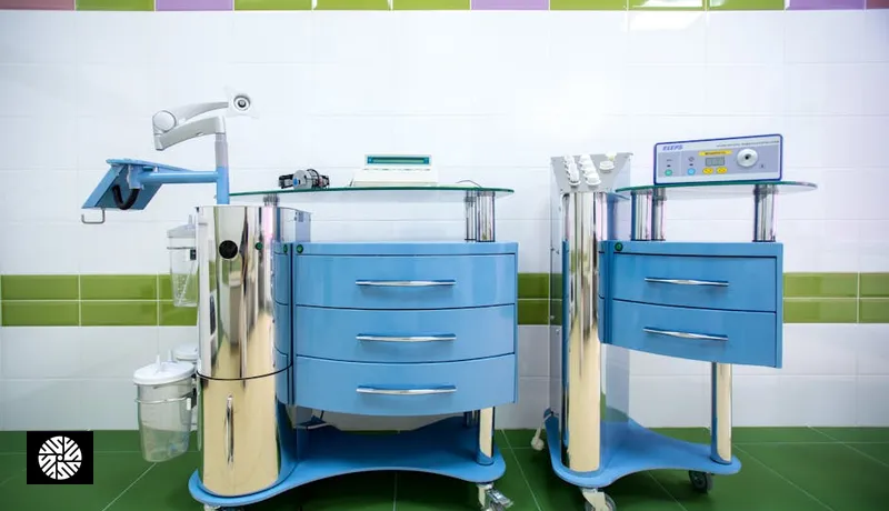
Women's preventive screening is not a smaller version of men's. It is a different protocol with different priorities — breast tissue, hormonal cycles, reproductive organs, bone density that drops sharply at menopause, and cardiovascular disease that has historically been underdiagnosed because the symptoms present differently. Taiwan's outpatient ecosystem makes the full panel — breast MRI on a 3T magnet, transvaginal ultrasound, DEXA scan, AMH and full hormone panel, gynecologic exam, and cardiac workup — accessible in a single coordinated visit, often inside a week, and at a fraction of US self-pay pricing.
This guide is the clinical-depth companion to our screening services overview. If you want the procedural side — what a full-body MRI day actually looks like — read our full-body MRI step-by-step guide first. For the cardiac angle that complements the women's protocol below, see heart and lung screening in Taiwan.
Women's screening — what differs from men's protocol
The men's preventive panel centers on cardiovascular disease, prostate, colon, and lung. The women's panel keeps cardiovascular and colon — those are universal — but adds four pillars men do not face: breast tissue surveillance, cervical and HPV screening, ovarian and pelvic evaluation, and a hormonal axis that shifts dramatically across reproductive years, perimenopause, and post-menopause. Bone density also matters earlier and more, because menopause-driven estrogen decline accelerates bone loss in a way men do not experience until later and more gradually.
The other structural difference is timing. Men can largely follow age-based triggers (CAC at 40, colonoscopy at 45, PSA discussion at 50). Women's screening is layered on top of a reproductive timeline — fertility window, contraception, pregnancy planning, perimenopause, post-menopause — and the right test set depends as much on life stage as on chronological age. A 35-year-old planning pregnancy and a 35-year-old with a BRCA-positive aunt are running very different protocols, even though their birth year is identical.
Breast cancer screening — mammography, DBT, MRI
Breast cancer is the most diagnosed cancer in US women — roughly 290,000 new invasive cases annually per ACS data — and the lifetime risk is approximately 1 in 8. The screening modalities have evolved meaningfully in the last decade, and the choice between them is no longer a single default for everyone.
Mammography is the baseline. The USPSTF carries a Grade B recommendation for biennial screening in women ages 50–74, and in 2024 expanded that to begin at age 40 — a notable shift after years of debate. Standard digital mammography uses low-dose X-ray to image compressed breast tissue and is highly effective for detecting microcalcifications, the earliest sign of ductal carcinoma in situ.
Digital breast tomosynthesis (DBT), often marketed as "3D mammogram," is now standard at most US academic centers and increasingly available in Taiwan. DBT acquires multiple low-dose projections across an arc and reconstructs thin slices, which improves cancer detection rates and reduces callbacks for benign findings, particularly in women with heterogeneously dense or extremely dense breast tissue (BI-RADS density categories C and D). For most women under 60, DBT — not 2D mammography alone — is now the preferred first-line screening modality.
Breast MRI is the highest-sensitivity modality and is reserved for high-risk screening. ACR guidelines support annual screening MRI for women with an estimated lifetime breast cancer risk of 20% or greater — which includes BRCA1/2 carriers, women with a strong family history (mother or sister with breast cancer, especially premenopausal), prior chest radiation between ages 10–30 (e.g., for Hodgkin lymphoma), and Li-Fraumeni or other inherited cancer syndromes. Breast MRI uses dynamic contrast-enhanced (DCE) protocols with gadolinium contrast. Image quality is meaningfully better at 3 Tesla than at 1.5T, which is why our Taiwan partners' 3T magnets matter — see Taiwan's 3T MRI standard for the technical reasoning.
| Modality | Best for | Sensitivity in dense tissue | Radiation | USPSTF / ACR position |
|---|---|---|---|---|
| 2D digital mammography | Baseline screening, microcalcifications | Reduced | Low-dose X-ray | USPSTF Grade B, 40–74 |
| DBT (3D mammogram) | Dense tissue, fewer callbacks | Improved over 2D | Slightly higher than 2D | Standard of care, US academic centers |
| Breast MRI (3T, DCE) | High-risk, BRCA, dense breasts cat. C/D | Highest of any modality | None (gadolinium contrast) | ACR: annual if lifetime risk ≥ 20% |
| Whole-breast ultrasound | Adjunct in dense breasts when MRI unavailable | Moderate adjunct value | None | Adjunct, not replacement |
Asian and Asian-American breast cancer specifics
The screening defaults written by US task forces are based on aggregate population data that historically underweighted Asian and Asian-American women. Two epidemiologic patterns matter for women in this group, and they push the recommendation toward earlier and more imaging-intensive screening.
First, age at presentation skews younger. Multiple registry studies — including SEER analyses of Asian-American subpopulations — show median age at breast cancer diagnosis 5 to 10 years earlier than non-Hispanic white women. The peak incidence in Asian populations clusters in the 40s and early 50s, while in white US populations it shifts later. A USPSTF "start at 40" guideline that already feels late for some patients can feel meaningfully late for an Asian-American woman with any family history.
Second, breast tissue is denser on average. Asian women are disproportionately represented in BI-RADS density categories C (heterogeneously dense) and D (extremely dense), and that pattern persists into the postmenopausal years where white populations tend to transition to fattier tissue. Dense tissue both raises baseline cancer risk and reduces the sensitivity of standard 2D mammography — the cancer hides in the white-on-white background. This is exactly the population in which DBT is more useful than 2D mammography, and where adjunct breast MRI or ultrasound carries the most additional value.
The practical implication: if you are an Asian or Asian-American woman, the conversation with your screening physician should explicitly address density and family history, and the default protocol — particularly between ages 35 and 50 — should bias toward DBT plus selective MRI rather than annual 2D mammography alone. Taiwan partners are accustomed to this population and routinely build that protocol without the patient having to advocate for it.
Cervical and HPV screening
Cervical cancer screening is the one area where US domestic preventive care is generally strong. USPSTF carries a Grade A recommendation for cervical cytology (Pap) every three years in women ages 21–65, or co-testing with HPV every five years for women 30–65. Most insured US women receive this consistently through their primary care or OB-GYN, and there is rarely an access gap that medical travel needs to fill.
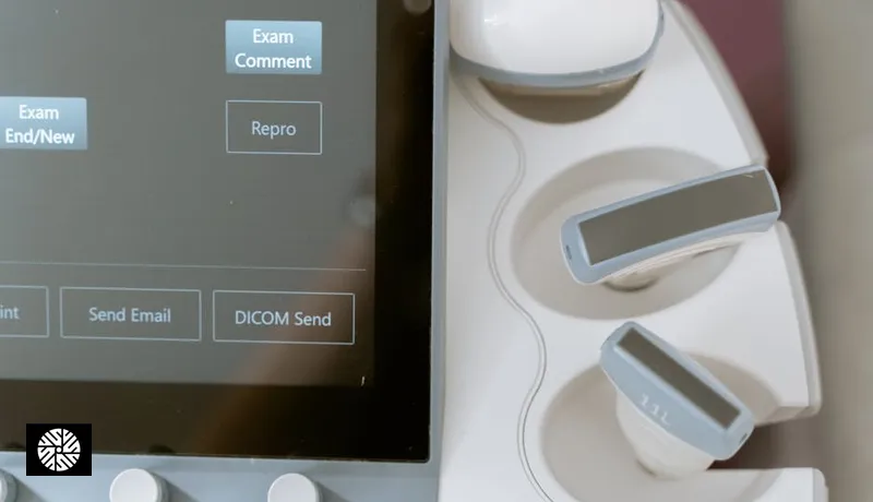
That said, our Taiwan partners offer integrated visits that bundle the gynecologic exam, Pap, HPV co-test, transvaginal ultrasound, and pelvic exam in a single appointment — useful if you are already coming for breast MRI or full-body imaging and would rather not run a separate OB-GYN visit at home. Results turnaround for cytology is typically 3–7 business days and is reported in English on request.
Ovarian and pelvic — when CA-125 + transvaginal ultrasound make sense
USPSTF carries a Grade D recommendation against routine ovarian cancer screening in average-risk asymptomatic women. The reasoning is that CA-125 alone has poor specificity (it elevates with endometriosis, fibroids, pregnancy, and many benign conditions) and transvaginal ultrasound alone has produced more harm than benefit in randomized trials of average-risk populations.
The story is different for the high-risk subset. Women with confirmed BRCA1 or BRCA2 mutations carry a lifetime ovarian cancer risk of roughly 40% (BRCA1) and 15% (BRCA2). Women with Lynch syndrome (mismatch repair gene mutations) carry roughly 10–12% lifetime ovarian and significantly elevated endometrial risk. For these patients, surveillance with CA-125 plus transvaginal ultrasound every 6 months is a reasonable bridge until risk-reducing salpingo-oophorectomy (typically recommended around age 35–40 for BRCA1, 40–45 for BRCA2). Strong family history without confirmed mutation — multiple first-degree relatives with ovarian, breast, or related cancers — also justifies the surveillance conversation.
If you fall into one of these categories and your US insurance is restrictive about repeat surveillance imaging, Taiwan's self-pay pricing makes the every-6-month protocol financially feasible. Our coordinators can also stage a pelvic MRI alongside breast MRI in the same visit.
Pelvic MRI for advanced screening
Beyond ovarian surveillance, pelvic MRI is a high-resolution tool for evaluating uterine fibroids, adenomyosis, endometrial thickness, ovarian morphology, and pelvic floor anatomy. It is the imaging modality of choice when a transvaginal ultrasound finds something ambiguous, when fibroid mapping is needed before considering myomectomy or uterine artery embolization, or when chronic pelvic pain has not been explained by simpler workup.
For perimenopausal women with abnormal bleeding, endometrial thickness on imaging matters: a thickened endometrium ( > 4mm in postmenopausal women, or asymmetric/heterogeneous appearance pre-menopausally) is the imaging trigger for endometrial biopsy. Pelvic MRI gives a more complete picture than ultrasound alone, particularly for staging if endometrial pathology is found.
DEXA bone density — establishing pre-menopausal baseline
USPSTF carries a Grade B recommendation for DEXA bone density screening in women age 65 and older, or in women under 65 with elevated fracture risk (low body weight, prior fragility fracture, long-term steroid use, family history). The Grade B 65+ default is well-known. What is less appreciated — and where the women's preventive panel adds value — is the case for a pre-menopausal or perimenopausal baseline DEXA.
Bone mineral density is relatively stable through reproductive years, then drops sharply in the 5–7 years around menopause as estrogen declines. Without a pre-menopausal baseline, you are measuring against a population reference rather than against your own peak bone mass. A baseline DEXA in your mid-to-late 40s — and certainly by age 50 if you have any risk factors (small frame, family history of osteoporosis, vitamin D insufficiency, restrictive diet history) — gives you a personal reference point. If a follow-up scan in your mid-50s shows a 12% drop, that is meaningful information you cannot extract from a single 65+ scan compared to population norms.
DEXA is fast (about 10 minutes), uses a fraction of the radiation of a chest X-ray, and at our Taiwan partners runs roughly 1/4 to 1/5 the US self-pay cost.
Hormone panel for women
The hormonal workup is where women's screening diverges most sharply from men's, and where the right test set depends almost entirely on life stage. The full panel includes estradiol (E2), follicle-stimulating hormone (FSH), luteinizing hormone (LH), anti-Müllerian hormone (AMH), thyroid panel (TSH, free T4, optionally free T3 and antibodies), and morning cortisol — with progesterone, prolactin, and DHEA-S added in specific contexts.
AMH deserves its own paragraph because it is the most useful single number for fertility window assessment. AMH reflects ovarian reserve — the size of the remaining follicle pool — and unlike FSH it is relatively stable across the menstrual cycle, so it can be drawn on any day. A 32-year-old planning to defer pregnancy by 5 years has very different decisions to make depending on whether her AMH is 3.5 ng/mL (reassuring) or 0.8 ng/mL (low for her age, worth a fertility consultation now). Taiwan partners can incorporate AMH into the screening visit and refer to fertility specialists if the result warrants.
| Life stage | Core hormone panel | Why it matters |
|---|---|---|
| Reproductive (20s–30s) | AMH, TSH, free T4, prolactin, vitamin D | Fertility window, thyroid baseline, cycle regulation |
| Pre-conception planning | Add FSH (day 3), LH, estradiol, progesterone (day 21), HbA1c, ferritin | Ovulation confirmation, anemia, glucose tolerance |
| Perimenopause (40s–early 50s) | FSH, LH, estradiol, AMH, TSH, free T4, AM cortisol | Cycle changes, mood, sleep, hot flushes workup |
| Post-menopause | FSH, estradiol, TSH, lipid panel, fasting glucose / HbA1c | Cardiometabolic shift, HRT decisions, thyroid drift |
Cardiovascular risk in women — historically underdiagnosed
Cardiovascular disease is the leading cause of death in US women, but for decades it has been underdiagnosed in women relative to men. The reasons are well-documented: symptom presentation differs (women more often present with fatigue, shortness of breath, jaw or back pain rather than the classic crushing chest pain), risk calculators were validated primarily in male cohorts, and microvascular disease — common in women — is often missed by standard angiography.
Two screening tools are particularly informative for women. Coronary artery calcium (CAC) score is a low-radiation chest CT that quantifies calcified plaque in coronary arteries and is among the strongest predictors of future cardiac events. A CAC of zero in a 50-year-old woman is genuinely reassuring; a CAC of 100+ in the same woman is a clear signal to escalate prevention regardless of how she feels. Lipoprotein(a) — Lp(a) — is a genetically determined lipid particle measured once in a lifetime. Roughly 1 in 5 people has elevated Lp(a), it is not modified by lifestyle or standard statin therapy, and it independently raises cardiovascular and aortic valve risk. It is particularly useful in women because traditional risk calculators (ASCVD, Framingham) tend to underestimate women's risk in the perimenopausal years where Lp(a)-driven risk is biologically active.
Both tests are bundled into our Taiwan partners' cardiac packages alongside ECG, echocardiogram, and stress testing where indicated. See heart and lung screening in Taiwan for the full cardiac protocol.
Patient personas — three concrete scenarios
The 35-year-old, fertility-focused. She is planning to start a family in 2–3 years and wants real data, not vague reassurance. Her panel: AMH plus a full reproductive hormone workup, transvaginal ultrasound to assess ovarian morphology and antral follicle count, thyroid panel, vitamin D and ferritin, HPV co-test, and a baseline breast ultrasound (DBT may be deferred if she has no family history and is comfortable waiting until 40). If AMH comes back lower than expected for her age, the visit ends with a fertility specialist referral — not a panic, just a recalibration of her timeline. Total cost is a fraction of equivalent self-pay in the US, and the visit fits inside a long weekend.
The 50-year-old, perimenopausal. Her cycles have become irregular over the last 18 months, sleep is disrupted, and she wants to make an informed decision about hormone therapy without flying blind. Her panel: FSH, LH, estradiol, AMH, full thyroid panel with antibodies, AM cortisol, DBT mammography, breast MRI (she has a maternal history of breast cancer at 58), transvaginal ultrasound, baseline DEXA, lipid panel, HbA1c, CAC score, and Lp(a). The result is a complete picture: where she sits in the menopause transition, whether her bones have started to drift, whether her cardiovascular risk justifies more aggressive lipid management, and whether HRT is a reasonable conversation. Her US insurance would have covered perhaps three of these tests. The Taiwan visit covers all of them in 4–5 days.
The 65-year-old, retirement-baseline. She is recently retired, generally healthy, and wants a comprehensive baseline before the next decade. Her panel: full-body MRI, DBT plus breast MRI (she has dense category-C tissue), transvaginal ultrasound, DEXA (now in the USPSTF-endorsed window), CAC score, Lp(a), full lipid panel, HbA1c, fasting glucose, thyroid, TSH, vitamin D, B12, kidney and liver function, and a colorectal screening discussion. The output is a structured longitudinal baseline she and her US primary care physician can refer back to for the next 10 years.
Bringing it together
The point of the women's preventive panel is not to do every test on every woman. It is to match the right tests to your life stage, your family history, and your specific risk profile — and to do that with imaging quality (3T MRI, modern DBT, current ultrasound) and turnaround that matches or exceeds US academic centers, at self-pay pricing that makes the comprehensive version actually achievable. For the broader case on why this works, read preventive health gaps in the US and how Taiwan fills the void.
Browse our screening providers in Taiwan to see which partners offer integrated women's panels, or contact New Dawn Health to scope a custom protocol for your specific risk factors and life stage. External references: USPSTF, American College of Radiology, American Cancer Society, CDC breast cancer, Taiwan NHI.
Sources & Further Reading
- USPSTF — U.S. Preventive Services Task Force recommendations
- American Cancer Society — Cancer Facts & Figures
- NCI SEER — Surveillance, Epidemiology, and End Results Program
- CDC — Cancer prevention & screening
- American Heart Association — heart attack risk
- American College of Cardiology — clinical guidelines
FAQ
USPSTF updated its guideline in 2024 to recommend biennial screening starting at age 40 (Grade B), through age 74. If you have a family history of breast cancer — especially a first-degree relative diagnosed pre-menopause — earlier screening is reasonable. For Asian and Asian-American women, the median age at presentation skews 5–10 years younger than non-Hispanic white women, which is another reason to have an explicit conversation about starting at 40 (or earlier) rather than waiting.
ACR guidelines recommend annual breast MRI in addition to mammography for women whose lifetime breast cancer risk is 20% or greater. That includes confirmed BRCA1/2 carriers, women with a strong family history (mother, sister, or multiple second-degree relatives with breast cancer), prior chest radiation between ages 10–30, Li-Fraumeni or related inherited cancer syndromes, and in some cases extremely dense breast tissue (BI-RADS category D) combined with other risk factors. If none of those apply, MRI is generally not recommended as routine screening.
AMH (anti-Müllerian hormone) is the single most useful number for assessing ovarian reserve and fertility window. It is most informative for women in their 20s to mid-30s who are deferring pregnancy and want to make informed timing decisions. AMH is relatively stable across the menstrual cycle, so it can be drawn on any day. A low result for your age does not mean you cannot conceive — it means a fertility specialist consultation is worthwhile sooner rather than later. AMH is not a useful test post-menopause.
USPSTF recommends DEXA at age 65, or earlier if you have specific risk factors. Beyond that, a baseline DEXA in your mid-to-late 40s (or by age 50) gives you a personal reference point before menopausal estrogen decline accelerates bone loss. Without a baseline, a single scan at 65 only tells you how you compare to population norms — not how much bone you have actually lost from your own peak. The scan is fast, low-radiation, and at Taiwan partners runs at roughly 1/4 the US self-pay cost.
CA-125 is not reliable as a routine screening test in average-risk asymptomatic women — USPSTF carries a Grade D recommendation against it. Specificity is poor because CA-125 elevates with endometriosis, fibroids, pregnancy, pelvic inflammation, and many benign conditions. The exception is the high-risk subset: confirmed BRCA1/2 carriers, Lynch syndrome carriers, or women with strong family history of ovarian or related cancers. In those patients, CA-125 paired with transvaginal ultrasound every 6 months is a reasonable surveillance protocol, typically as a bridge to risk-reducing surgery.
HPV vaccination dramatically reduces but does not eliminate cervical cancer risk — current vaccines cover the highest-risk strains (16, 18, and several others) but not every oncogenic HPV type. USPSTF Grade A cervical screening recommendations apply regardless of vaccination status: cytology every 3 years from 21–65, or HPV co-testing every 5 years from 30–65. Vaccination is a complement to screening, not a replacement.
Yes — the typical women's panel (breast imaging including MRI, transvaginal ultrasound, gynecologic exam with Pap and HPV, DEXA, full hormone panel including AMH, cardiovascular workup with CAC and Lp(a), and a comprehensive blood panel) fits inside 4–5 working days. Image-heavy results (MRI, DEXA, ultrasound) are typically read within 1–2 days; lab work including AMH and full hormone panel turns around in 2–3 days. English reports are standard at our partner clinics. Contact our coordinators to scope your specific protocol before booking.