What is a comprehensive eye exam?
A comprehensive eye exam checks both how well you see and how healthy your eyes are. Beyond measuring visual acuity and refraction (the prescription for glasses), it usually includes a dilated look at the retina and optic nerve at the back of the eye. Many eye diseases cause no symptoms early on, which is why the look inside matters. It is painless; the main inconvenience is a few hours of blurred vision and light sensitivity after the drops.
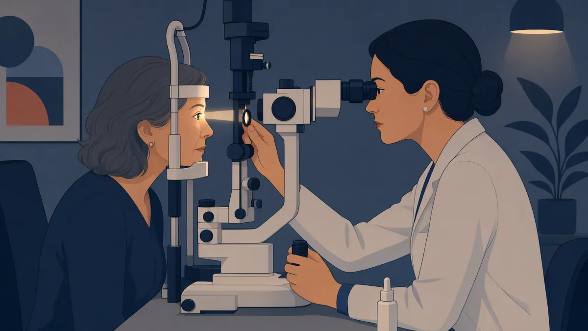
How the exam works, and why the drops
The pupil is not a structure; it is an opening — the round hole in the iris, the colored ring. The iris is a muscle that changes the size of that hole: wide in dim light, narrow in bright light to protect the retina. For this exam that reflex is a problem. To see the back of the eye, the examiner shines a bright light in through the pupil, and the iris closes the very hole they are trying to look through.
Dilating drops hold the hole open against the light for a few hours. The NEI compares it to opening a door to let light into a dark room.
Why the eye is worth this trouble
Light goes into the eye through a clear window and comes back out. So the retina, the light-sensing layer at the back, can be looked at directly in a living person with nothing cut open. Small blood vessels lie on its surface, and at one spot the optic nerve leaves the eye toward the brain.
That makes the retina one of the few places where blood vessels and a nerve can be seen directly, with no needle and no scan. Diabetes and high blood pressure damage small blood vessels throughout the body, and here that damage can sometimes be seen. Seeing damage is not the same as an early diagnosis: retinal changes usually follow years of raised blood sugar or pressure.
What an eye exam can find
A dilated eye exam looks for the common causes of vision loss, several of which are silent at first:
- Glaucoma — slow optic-nerve damage that often has no early symptoms
- Age-related macular degeneration — a cause of central vision loss with age
- Cataracts — clouding of the lens that gradually dims and blurs vision
- Diabetic retinopathy — vessel damage in people with diabetes, often before vision changes
- Blood-vessel changes from high blood pressure
The last two are signs, not tests. Diabetes is diagnosed with blood tests such as HbA1c or fasting glucose, and high blood pressure with a blood-pressure cuff.
What an eye exam does not cover
- How sharply you see, and your glasses prescription
- The front of the eye, the lens, the retina and the start of the optic nerve
- Whether you have diabetes. A blood panel with HbA1c or fasting glucose answers that.
- Your blood pressure. A cuff measurement does, as in a physical exam.
- The socket and the optic nerve behind the eye. When symptoms or exam findings point there, a doctor orders a dedicated MRI of the orbits and brain. A survey full-body MRI is not built for that question.
Where an eye exam fits in a screening
How often you need one depends on your age and risk. US guidance from the National Eye Institute and the American Academy of Ophthalmology:
| Who | How often | Source |
|---|---|---|
| Healthy adults under 40 | Every 5 to 10 years, with a baseline exam by 40 | AAO |
| Between 40 and 64 | Ask your eye doctor, who sets the interval after the baseline exam | AAO |
| Over 60, African American over 40, or family history of glaucoma | A dilated exam every 1 to 2 years | NEI |
| 65 and older | Every year or two | AAO |
| Diabetes or high blood pressure | Most people at least once a year; ask your doctor | NEI |
At New Dawn Health in Taipei an eye exam may sit in a package alongside a blood panel. Who performs it, whether dilation is included and the report language are confirmed when you book. Take the result to your own eye doctor so it joins your record.