What is an MRI?
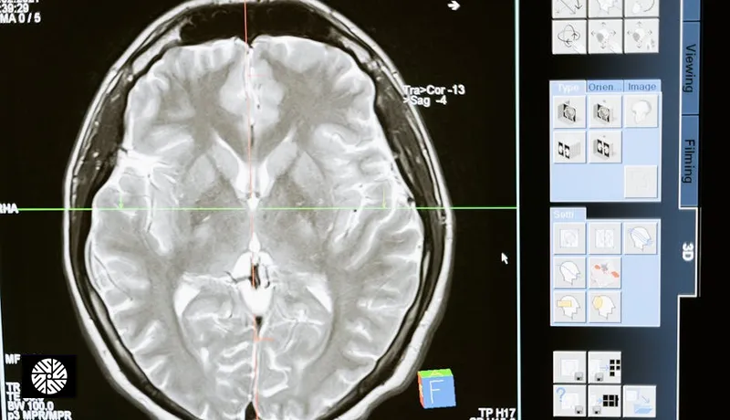
Magnetic resonance imaging — MRI — builds detailed cross-sectional pictures of the inside of your body using a powerful magnet and radio waves. It shows soft tissue in fine detail: brain, organs, discs, muscle, and the boundary between a tumor and the healthy tissue around it.
An MRI uses no ionizing radiation. There is no X-ray beam in the machine, so no radiation dose accumulates from one scan to the next. That removes one harm. It does not, on its own, make scanning a healthy person useful; the section on uses below says where the evidence stands.
How an MRI actually works
The physics has a reputation for being impenetrable. It is not. Your body is mostly water, and every water molecule contains hydrogen. The nucleus of a hydrogen atom is a single spinning proton, and a spinning charge behaves like a tiny magnet. Normally those magnets point every which way, so they cancel out. Inside the scanner, a strong magnetic field pulls them into line. For the first time they add up instead of canceling.
The scanner then sends a burst of radio energy at exactly the frequency those protons respond to, and it tips them out of line. When the pulse stops, they drift back and release the energy as a faint radio signal, which coils around your body pick up. Different tissues settle back at different speeds — and that difference is the picture. The scanner is not measuring what a tissue is. It measures how quickly that tissue's hydrogen recovers, and turns the timing differences into light and dark.
| Sequence type | What it makes bright or dark | Why a radiologist wants it |
|---|---|---|
| Fluid-sensitive | Water glows | Cysts and swelling become obvious |
| Fat-suppressed | Fat is turned dark | An inflamed area is not hidden behind fat |
| Flow-sensitive | Moving blood stands out | Maps arteries, as in an MR angiogram |
A sequence is one of those recipes: a pattern of radio pulses, timed to make one kind of tissue stand out from its neighbors. A scan runs the same body through several of them, and the radiologist reads them together. That is why an MRI takes much longer than a CT.
Why it is so loud
An MRI knocks, taps and hammers loudly enough to sound as if it is breaking. It is not. The returning radio signal carries no return address: every proton sings the same note. So the scanner makes the field deliberately uneven — it briefly tilts the field in different directions — because a proton's note depends on the field where it sits. Now position and pitch are linked, and the scanner can work out where each signal came from. The gradient coils that tilt the field switch on and off very rapidly. Each time a strong current runs through a wire inside a huge magnet, the wire is shoved sideways and the coils flex against their mountings. You are hearing the machine physically vibrate as it works out where you are.
Why they keep asking about metal
The magnet is always on. It is not switched on for your scan and off afterwards. In the usual superconducting scanner the main magnet stays on, including while the room sits empty and silent. A scanner that looks idle and harmless is at full field, and the signs on the door are the only warning.
That creates two separate dangers. Anything the magnet can grab — scissors, keys, an oxygen cylinder — is snatched toward the tunnel, where a person's head is. And metal inside you can absorb radio energy and heat up, or a device's electronics can be disturbed by the field. The items that matter most:
- Pacemakers and implanted defibrillators, and other implanted stimulators
- Cochlear implants, some of which contain a magnet of their own
- Some aneurysm clips — an older ferromagnetic clip on a brain artery is a genuine contraindication
- Metal fragments — a bullet, shrapnel, or a splinter in the eye from grinding or welding metal
Many modern implants are safe in an MRI, and some pacemakers are designed for it. But "safe" depends on the exact model, and often on the scan settings. A single "any metal?" question cannot settle that, which is why screening is item by item and may be checked more than once.
What else to tell the team before the scan
- If you are or could be pregnant. RadiologyInfo says there is no known risk from MRI in pregnancy, but a non-urgent scan may be rescheduled. Most centers avoid contrast in pregnancy.
- If you are breastfeeding and contrast is planned. RadiologyInfo says there is no need to interrupt breastfeeding; a parent who prefers a short pause can ask the team.
- Kidney or liver disease, or a past reaction to MRI contrast, if contrast is planned.
- Claustrophobia — before you book, not on the day.
Claustrophobia — the honest section
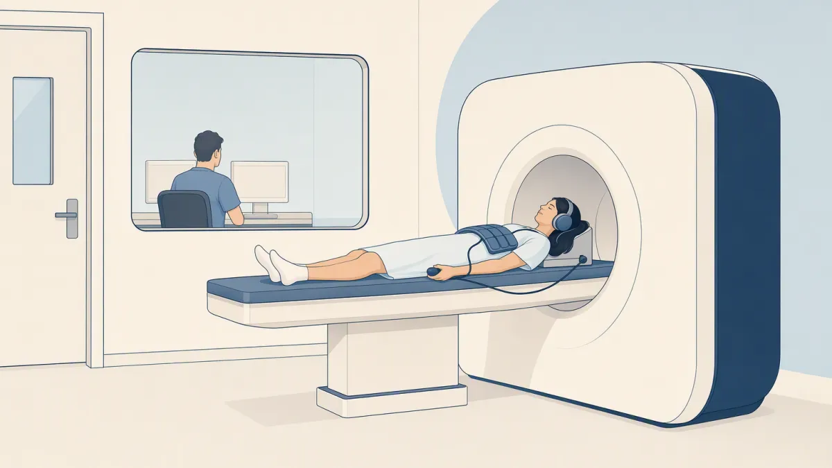
An MRI means lying still inside a tunnel, often for a long time, while it hammers. Your coordinator confirms the expected duration of your protocol when you book. A typical closed-bore scanner is a cylinder open at both ends; designs vary by site. For a head or chest scan your face sits inside it.
Many people tolerate it easily; a real minority do not. RadiologyInfo notes that about one person in twenty may need a mild sedative to stay calm. That is a normal response to an abnormal situation, not a measure of how tough you are. Things that can help:
- Knowing the shape of the scan in advance
- A feet-first position where the anatomy allows, so your head stays outside the bore
- Music, an eye mask, and talking to the technologist between sequences
- A wider or more open scanner — not every site has one, so ask before you book
RadiologyInfo says that with good preparation it is almost always possible to complete the exam. Almost is the honest word. Some people still cannot finish, and that is a limit of the machine, not a personal failing. It is worth raising before you have paid for a scan.
Contrast — will I be injected?
It depends on the protocol. Many MRI scans run with no contrast at all. Breast MRI uses it, and an MR angiogram may be done with or without it. Your coordinator confirms what your protocol involves when you book.
Because the picture is shaped by blood flow, contrast is used where blood supply is the question. A growing tumor recruits its own vessels, which is why breast MRI relies on it. Retained traces are one reason contrast is given when it answers a question, not by default.
What MRI is used for
Most MRI is diagnostic: it answers a question raised by symptoms or an earlier finding. As a screen for people who feel well, its role is narrower than the technology suggests. In this wiki MRI fans out into three exams, each with its own page:
| Exam | What MRI is asked to show | Who it is for |
|---|---|---|
| Full-body MRI | A survey of several regions; coverage varies by protocol | Not routinely recommended for average-risk adults without symptoms (ACR, 2023) |
| Brain MR angiogram | Blood vessels in the head, with or without contrast | Usually a clinical indication or selected higher-risk people |
| Breast MRI | Blood-flow patterns in breast tissue, with contrast | People at high risk, in addition to mammography (American Cancer Society) |
Where MRI falls short
Most of MRI's blind spots come straight out of the physics above. It reads hydrogen in water and fat, so wherever there is little mobile hydrogen — water or fat — there is little picture.
- Soft tissue and solid organs — brain, liver, kidneys, prostate, uterus
- Masses, distinguished from the tissue around them
- Blood vessels, sometimes with no injection at all
- Spinal discs and the spinal cord beside them
- Fluid — cysts, swelling, inflammation
- Lungs — mostly air, so almost no hydrogen to signal; a low-dose lung CT is the tested screen for people who qualify
- Calcified plaque — hard calcium holds no mobile hydrogen; a heart calcium score measures it by density instead
- Colon polyps — a colonoscopy can find many polyps and remove them in the same procedure
Air and dense bone are where conventional MRI is weakest. A lung is essentially a bag of air, and air has almost no hydrogen in it, so there is nothing to sing back. The boundary between air and tissue also distorts the local field and scrambles the signal near it. Dense bone and calcium fail differently: they hold very little mobile hydrogen, and their signal fades too fast to catch. So lungs and coronary calcium go to CT.
Motion is the practical limit. An MRI image is assembled from signal collected over minutes, so anything that moves during those minutes smears. That is why you are asked to lie still, and why some sequences ask for a breath-hold of a few seconds.