What is comprehensive breast imaging?
Breast imaging can mean a mammogram, a breast ultrasound, a breast MRI, or a combination. They are not automatically combined for everyone. Which test fits depends on your age, your risk, your breast density, any symptoms and any earlier findings.
Mammography remains the main screening test for most women. In Taiwan the Health Promotion Administration funds a mammogram every two years for women aged 40 to 74.
How the three tests differ
Ultrasound and MRI both use no ionising radiation; mammography does. Each test sees the breast in a different way:
| Test | How it works | Main strength | Main trade-off |
|---|---|---|---|
| Mammogram | Low-dose X-ray, about 0.28 mSv for a screening mammogram | Shows microcalcifications, tiny calcium specks that can be an early sign of some cancers; screening programs are built on it | Less sensitive in dense breast tissue |
| Breast ultrasound | Sound waves; no radiation | Tells solid lumps from fluid-filled cysts and reads dense tissue | Used for screening, it finds many abnormalities that turn out not to be cancer |
| Breast MRI | Magnet and radio waves, usually with a contrast injection; no radiation | The most sensitive of the three; used for people at high risk | More false alarms and extra biopsies; not for average-risk screening |
Who needs which test
Pregnancy and breastfeeding change which test is used, so tell the team about either before booking.
| Situation | Usual approach |
|---|---|
| Women 40–74 at average risk | A mammogram every two years. Taiwan funds this; the US Preventive Services Task Force recommends the same. |
| Dense breasts on a previous mammogram | Mammography continues. Adding ultrasound or MRI is a discussion with your doctor, not a rule. |
| High risk | The American Cancer Society (US) advises a yearly MRI plus a mammogram, typically from age 30. This covers a lifetime risk of about 20–25% or more, or a known pathogenic or likely pathogenic BRCA1 or BRCA2 variant. |
| Pregnant or breastfeeding | In pregnancy, ultrasound is often the first test for a lump because it uses no radiation; contrast MRI is generally avoided. Breastfeeding alone does not replace an indicated mammogram. |
| A lump, nipple discharge or skin change | This is not screening. See a doctor, who will order diagnostic imaging for that symptom. |
What it can't tell you
Screening finds suspicious areas; it does not diagnose them. A suspicious area needs more imaging or a biopsy before anyone can say what it is. The main limits:
- False alarms are common: RadiologyInfo puts recall for more testing at 5–15% of screening mammograms. Most of those tests turn out normal.
- Over a decade they add up: with yearly mammograms from 40 to 49, the chance of at least one false positive is about 30%, and of a biopsy about 7–8%.
- MRI adds sensitivity and false alarms together. A suspicious area seen only on MRI can still need a targeted ultrasound or an MRI-guided biopsy, even when the mammogram and ultrasound were clear.
- A normal result is a snapshot. Screening works as a repeated interval, not a one-off.
- It covers the breasts only. Cervical screening is a separate test — see the gynecology exam.
What to expect on the day
-
1Safety and history firstTell the team if you are or could be pregnant, or are breastfeeding. Before any MRI you also go through implants and metal history, and — if contrast is planned — kidney function and any earlier contrast reaction.
-
2Check in & changeGown on, valuables in a locker. Skip deodorant, powder and lotion that morning — they can show up on the mammogram as calcium-like spots.
-
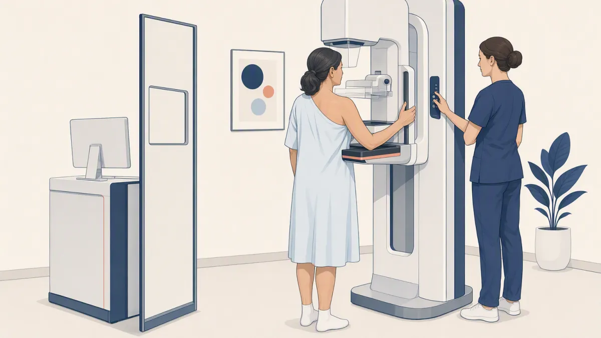
3Mammogram — seconds per viewEach breast is pressed between two plates for a few seconds per image. Some women find the compression painful; tell the technologist, who can adjust it.
-
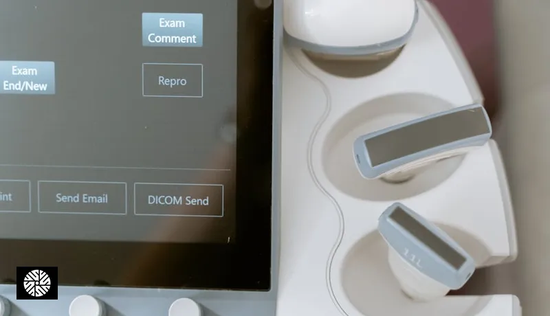
4Ultrasound, if it is part of your planWarm gel and a handheld probe sweep the breast. You feel light pressure.
-
5Breast MRI, if it is part of your planYou lie face down on a padded coil while the scanner images both breasts, usually with contrast through an IV line. It is loud, and you are given hearing protection.
Which tests your booking includes, their order, how long they take, and whether the MRI uses contrast are all confirmed by your coordinator when you book.
Reading your BI-RADS result
A breast imaging report ends with a single number between 0 and 6. That is the BI-RADS assessment category, and it is not a diagnosis. It is an instruction: the number says what the radiologist wants done next.
A ladder like this is the general pattern, not a rule your case must obey. Reporting conventions differ between hospitals, and two experienced clinicians can read the same finding and reasonably advise differently. Where your report or your doctor differs from this table, go with them — they are looking at your actual result.
These categories are the American College of Radiology standard, so a doctor at home can act on the number from a report issued in Taipei. Ask for the report language you need when you book.
When more than one test was done
If you had more than one test, the radiologist may read them together and give one category for the case. Your coordinator confirms whether your clinic reports this way. A combined read matters most when the tests disagree:
- Ultrasound can settle a mammogram question. A shadow with no clear shape on a mammogram may need more work on its own. An ultrasound of the same spot may show a simple fluid-filled cyst — a category 2 finding.
- It does not work in reverse for MRI. In other words, a clear mammogram and ultrasound do not cancel a suspicious MRI finding.
Cost and coverage
The live price further down this page is the booking price at New Dawn Health; anything billed separately — such as biopsy or pathology where it applies — is confirmed when you book. What happens after that depends on where your coverage comes from:
- Taiwan's funded mammogram is for eligible residents. The Health Promotion Administration program covers women 40–74 in the national system; visitors' screening in Taiwan is self-funded.
- Reimbursement at home is a question for your insurer. Whether a foreign insurance plan or health savings account pays any of it back depends on that plan, and on whether the test was screening or a follow-up.
Results and follow-up
- A written report with a BI-RADS category — a standard breast specialists elsewhere use.
- Turnaround and who explains it vary by hospital. Your coordinator confirms both when you book.
- A next step. Usually your routine interval, a short-interval follow-up, or a referral for more imaging or a biopsy that you can take to your own doctor.