What is an electrocardiogram (ECG)?
An electrocardiogram — ECG, sometimes written EKG — records the electrical signals that make your heart beat. Small, sticky electrodes on your chest, arms and legs read the tiny voltage changes of each beat, and the machine traces them as the familiar line of peaks. It is painless, uses no needles and no radiation, and takes a few minutes.
How an ECG works
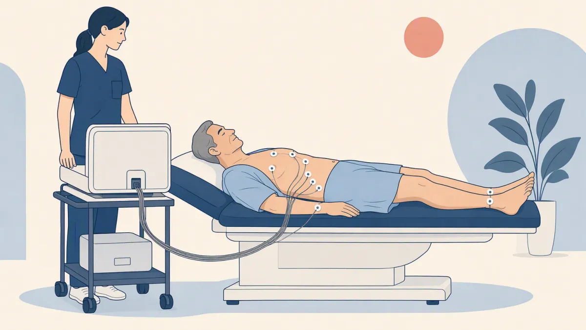
Every heartbeat starts as electricity. A small patch of cells near the top of the heart fires on its own, and that spark spreads as a wave from cell to cell. Muscle contracts where the wave arrives, so the squeeze follows the wave in order — which is what makes a heart a pump. You are mostly salt water, and salt water conducts, so the wave is faintly detectable at your skin. The electrodes send nothing into you; they only read the voltage underneath them.
Here is why there are several of them. An electrode pair only sees the part of the wave heading its way: a wave traveling toward it traces upward, a wave traveling away traces downward. Each pair — each lead — watches the same wave from a different angle. Put the viewpoints together and the tracing shows the path the wave took through the heart, and where it struggled to cross.
What it is like
-
1Electrodes onSmall sticky electrodes go on your chest, arms and legs. A patch of chest hair may need shaving so they stay on.
-
2Lie stillThe machine records while you lie quietly. You feel nothing, because nothing enters your body.
-
3Electrodes offThe whole test takes a few minutes. Some people notice mild skin irritation where the patches were.
What the peaks actually are
The familiar shape is three events in a fixed order:
- P — the wave crossing the two small upper chambers, which collect blood. Little muscle, so a small bump.
- QRS — the wave sweeping through the two large lower chambers that drive blood out to the body. Most of the heart's muscle is here, so this is the tall spike.
- T — the lower chambers resetting electrically so they can fire again for the next beat.
The flat stretches between are not nothing happening; they are nothing moving. Rhythm problems show up as this sequence arriving early, late, out of order, or not at all.
What an ECG can find
An ECG reads your heart's rhythm and rate, and from that alone it can flag:
- Atrial fibrillation — an irregular rhythm that may cause no symptoms and raises the risk of stroke
- A previous heart attack, sometimes one that went unnoticed
- A heartbeat that is too fast or too slow, or a conduction problem
- Indirect clues to a thickened or enlarged heart muscle
What an ECG can't tell you
A resting ECG says nothing about the plaque inside your arteries. Electricity is not plumbing. The electrodes read the wave traveling through heart muscle; the coronary arteries are the pipes delivering blood to that muscle, and they cast no signal of their own. The tracing mainly changes when the muscle has been damaged, or is short of blood at the moment of the recording.
A narrowed artery at rest may be doing neither. The muscle it feeds still gets enough blood to conduct normally, so the wave crosses it normally and the ECG has nothing to report. That is why a resting tracing can be completely normal in someone with significant coronary disease.
- Your rhythm and rate during the recording
- Signs of past damage to the heart muscle
- A rhythm problem that comes and goes. An ECG is a snapshot; a portable monitor worn for a day or longer can catch it.
- Calcified plaque. A heart calcium score measures it, used selectively when a prevention decision is uncertain.
- How narrow an artery is. A coronary CT angiogram shows the narrowing; a stress test shows whether it limits blood flow. Both answer a defined clinical question.
Where the ECG fits in screening
The ECG answers one narrow question — is the rhythm normal right now? — not the broader one most people have in mind, are my arteries clear? How US guidance treats it as a screen:
- Low risk, no symptoms: the US Preventive Services Task Force (2018) recommends against resting or exercise ECG screening.
- Intermediate or high risk, no symptoms: it finds the evidence insufficient to judge the balance of benefits and harms.
- An abnormal tracing: a doctor decides the next step, such as a repeat ECG, a portable monitor, or a cardiologist referral.
- Symptoms: an ECG is a diagnostic test, and symptoms need a doctor's assessment rather than a screening package.
Heart-attack risk itself rests on blood pressure, cholesterol, diabetes and smoking, with family history weighed alongside. A blood panel covers the laboratory part. A normal ECG should not automatically lead to a calcium score or a CT angiogram.