What is an X-ray?
An X-ray is the oldest and simplest form of medical imaging. A quick, low-dose beam passes through the body and casts a flat shadow of your densest structures — mainly bone — onto a detector. It takes seconds and needs no preparation, which is why it has stayed in use for more than a century. It can be a diagnostic building block in a package, but for screening a healthy adult a plain X-ray has no established role.
How an X-ray works
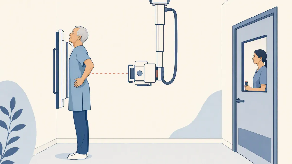
X-rays are the same kind of thing as visible light, but each ray carries far more energy, so they pass through material your eyes cannot see through. How easily they get through depends on what is in the way. Soft tissue is mostly water and fat, built from light atoms, and X-rays slip through it. Bone, packed with calcium, stops them, as do metal implants. The detector records how many X-rays arrived at each point, so the image is a map of what stopped the beam.
The display copies how photographic film behaved. Film darkened wherever X-rays struck it, so the air-filled lungs, which stop almost nothing, read black. Bone blocks the beam, so the area behind it stays white, and soft tissue comes out in shades of gray.
| What the beam passes through | How much it stops | How it looks |
|---|---|---|
| Air in the lungs | Almost nothing | Black |
| Soft tissue and fluid | Some | Shades of gray |
| Bone and metal | Most | White |
Why everything ends up flattened
A shadow has no depth. The beam travels in one direction, and everything it passes through — ribs, heart, lungs, spine — is summed onto one flat plane. Two structures at different depths cannot be separated, because their shadows land on top of each other. That is why a small lung nodule can hide behind a rib, and why an X-ray answers "is the bone broken?" far better than "is there something small in there?"
A CT scan uses the same physics to solve that limit. It takes hundreds of shadows from all around you, and a computer works out which arrangement of tissue would cast all of them, recovering the depth one shadow threw away.
What you experience, and the dose
-
1PositioningYou stand or lie against the detector and the technologist positions you. Remove jewelry and anything metal over the area.
-
2A held breathFor a chest film you hold your breath for a moment so the lungs stay still.
-
3The exposureIt lasts a fraction of a second. There is no heat, no sound from the beam, and nothing to recover from.
RadiologyInfo puts an adult chest X-ray at about 0.1 mSv (RadiologyInfo figures, checked October 2026). On its own that number means little, so anchor it: the average person in the US receives about 3 mSv a year from natural sources. A chest film is therefore about 10 days of average US background radiation. A coast-to-coast US round-trip flight adds about 0.03 mSv — about a third of a chest X-ray.
What an X-ray shows well
An X-ray reads dense structures best, and bone most reliably of all. It is the first-line test for a suspected fracture, and a chest X-ray can show large, obvious changes. What it cannot do is separate the overlapping soft tissues that screening usually cares about.
- Bone fractures and alignment
- Large chest changes — pneumonia, a collapsed lung, fluid, an enlarged heart shadow, a large mass
- The position of implants, tubes and hardware
- Early lung cancer — a low-dose lung CT, for people who meet the criteria
- Organ and soft-tissue detail — an MRI or an abdominal ultrasound, when there is a question to answer
- Bone density — a DEXA scan, itself a specialized very-low-dose X-ray
Where a better scan is warranted
The flat, single-plane picture stacks organs on top of one another, so small or soft-tissue changes are easily hidden. That is why lung-cancer screening does not use it.
Where a plain X-ray fits in screening
A plain X-ray fits wherever a fast answer about bone or obvious chest changes is enough. At New Dawn Health it may appear as one component of a package. When the goal is early detection of a specific disease, the scan built for that job is the one to ask about:
- Lung cancer — low-dose lung CT, if you meet the criteria
- Osteoporosis — a DEXA scan, by age, sex and fracture risk
- A finding on the film — usually a CT or another targeted test, chosen by the doctor who reads it