The radiation-free first look — high-frequency sound waves that image the thyroid, abdomen and arteries in real time, with no X-rays or needles.
Last updated October 2026 · New Dawn Health editorial team
How it works
High-frequency sound waves, real-time images
Radiation
None — no X-rays, no needles
Prep
Depends on the area; confirmed when booking
Where it fits
A radiation-free look for a defined question
What is an ultrasound?
An ultrasound is a radiation-free imaging test that uses high-frequency sound waves to picture soft
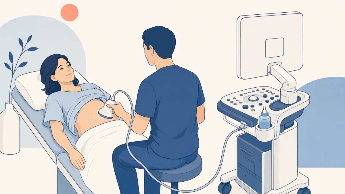
tissue in real time — no X-rays, no needles, no dye. A sonographer glides a handheld probe over the
skin and a live image appears on screen. Because it is quick and comfortable, it is often the first
look a doctor orders when there is a question about an organ, a lump or blood flow.
The image forms under the operator's hand, so ultrasound is a skill as much as a machine.
How ultrasound works
The probe sends pulses of high-frequency sound into the body and listens for the echoes that bounce
back off tissue boundaries. Different tissues reflect sound differently, and the scanner turns those
echoes into a moving grayscale image. Because it captures motion live, it can show a beating heart
valve, blood flowing through an artery (Doppler mode), or an organ shifting as you breathe.
How common materials look on an ultrasound.
What the sound meets
What happens to it
How it looks
Fluid (a cyst, a full bladder)
Passes through with few echoes
Dark
Soft tissue and organs
Returns a mix of echoes
Shades of gray
Bone, stones, gas
Reflected or scattered almost entirely
Bright, with a shadow behind
There is no ionizing radiation. The FDA notes that ultrasound energy can still warm tissue slightly,
so it is used prudently, when there is a medical reason, rather than repeated without one.
What it can show
In this wiki ultrasound fans out into three exams. Being able to see an organ is not the same as
screening it being worthwhile, and US guidance treats them differently:
The three ultrasound exams on this site and their screening role.
Not recommended routinely (USPSTF, 2021); used after symptoms or another indication
Where ultrasound falls short
Ultrasound cannot see through bone or gas, and it is only as good as the person holding the probe.
Sound reflects off the skull and scatters in air, so the brain, the lungs and anything behind bowel
gas are effectively invisible. Image quality also varies with the sonographer's skill.
✓
Reads well
Fluid-filled structures, solid organs near the surface, and moving blood
The same area again later, with no radiation
!
Hands off to another test
The brain behind the skull — an MRI, when there is a clinical question
The lungs — a low-dose lung CT for people who meet the screening criteria
Organs hidden by bowel gas, such as parts of the pancreas — a CT scan or MRI, chosen by the doctor
Ultrasound uses sound waves rather than ionizing radiation, and there is no injection. The FDA notes that the energy can warm tissue slightly, so it is used prudently, for a medical reason. You may feel light pressure from the probe and cool gel on the skin.
What can an ultrasound not see?
+
Ultrasound cannot see through bone or gas. Sound reflects off the skull and scatters in air, so the brain, lungs and structures behind bowel gas stay out of view. For those, a doctor may choose MRI or CT instead.
Do I need to prepare for an ultrasound?
+
It depends on the area. Thyroid and carotid scans usually need none. An abdominal ultrasound often asks you to fast for some hours so the gallbladder is full and bowel gas is reduced. Your coordinator confirms the rules when you book.
Is a thyroid or carotid ultrasound a good screening test?
+
Not for adults without symptoms. The US Preventive Services Task Force recommends against routine screening with either. Both are useful when there is a neck lump, symptoms, or another reason a doctor identifies.