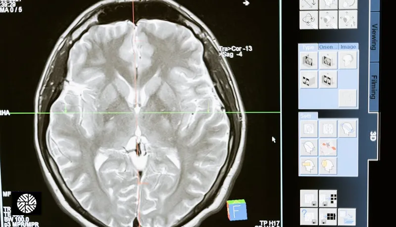
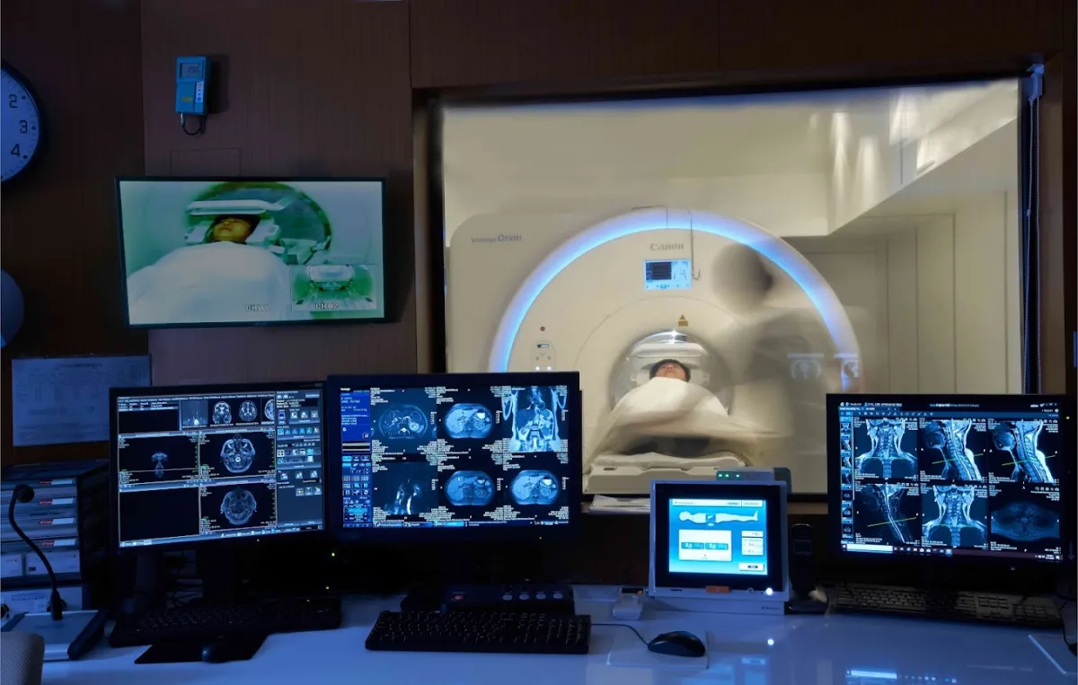
MRI uses no radiation, but that alone is not a reason to scan. A head scan is most useful when it answers a defined question — a symptom, an earlier finding or a known higher risk.
What gets checked, and with what
| Organ | What imaging looks for | The test that does it |
|---|---|---|
| Brain | Tumors, silent small strokes, atrophy (shrinkage of brain tissue) — and, on the artery sequence, unruptured aneurysms | Full-body MRI, when the protocol covers the head, plus a brain MR angiogram for the arteries |
| Eye | A mass or optic-nerve change behind the eye | A dedicated MRI ordered for symptoms; a full-body MRI may incidentally show the orbit |
| Ear | A tumor on the hearing nerve, such as an acoustic neuroma | A dedicated MRI ordered for symptoms; a full-body MRI may incidentally show the inner ear |
| Sinus | Chronic inflammation — thickened lining or trapped fluid | Full-body MRI, when the protocol covers the head |
Brain
- Why it matters: silent small strokes and most unruptured aneurysms cause no symptoms, so they are usually found on a scan done for another reason.
- Two different pictures: MRI shows the substance of the brain; MR angiography shows its blood vessels. Neither uses radiation. An MRA may be done with or without a contrast injection, depending on the protocol.
- Who the artery scan is for: people with symptoms, an earlier finding or a defined higher risk. The brain MR angiogram page sets out which family histories and conditions count.
- Imaging can't: test how the brain works. Memory, thinking and reflexes are assessed clinically — a clean scan is not a normal neurological exam.
Eye
- Why it matters: the orbit (the eye socket) and optic nerve sit inside the field a head MRI often covers, so a structural problem can be seen without a separate scan.
- Imaging can't: tell you how well you see. Vision, eye pressure and the retina belong to a dedicated eye exam.
Ear
- Why it matters: MRI is the test that images the hearing nerve, where a small benign tumor can grow slowly without being noticed.
- Imaging can't: measure hearing. That needs an audiogram and a look inside the canal — an ENT exam. Hearing loss on one side needs that testing whatever the scan shows.
Sinus
- Most common finding: thickened lining or a little fluid, often noted in passing on head scans.
- Imaging can't: tell you whether it matters. Sinus images correlate poorly with symptoms; that call belongs to an ENT physician, not the scan.
What this region's screening does not cover
What a head scan can give you
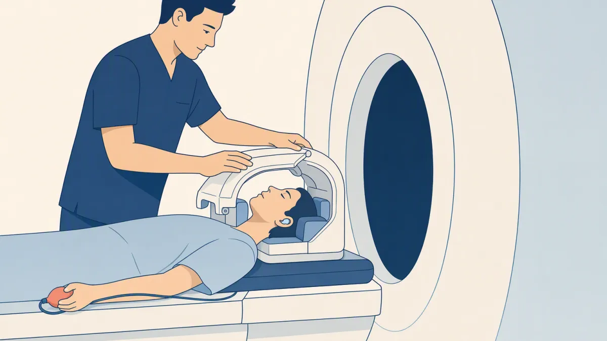
- Four structures in one pass, with no radiation, when the protocol covers the whole head
- The arteries too, if an MRA sequence is added
- A baseline to compare against if anything changes later
What it does not
- Function — vision and hearing are tested at an eye exam and an ENT exam, and thinking by a neurologist
- A guarantee. A normal MRA cannot exclude every future stroke or vascular event, and very small aneurysms can be missed.
- Incidental findings are common here: small cysts, benign variants, minor sinus thickening. They are usually monitored rather than treated, and a note on the report does not mean something is wrong.