What is a brain MR angiogram?
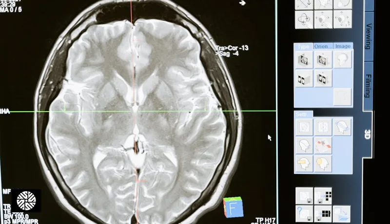
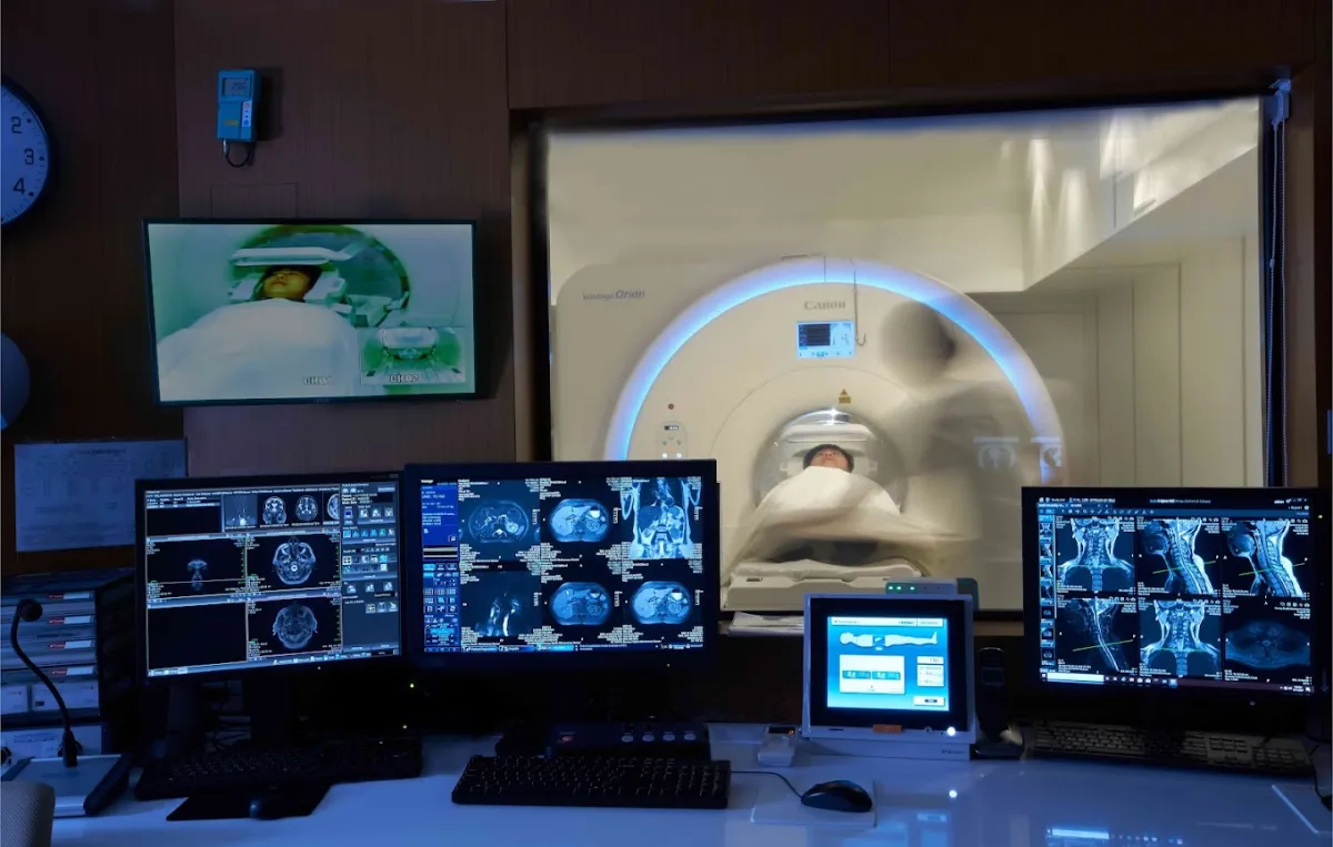
A brain MR angiogram (MRA) is an MRI scan tuned to show the blood vessels inside your head rather than the brain tissue. It uses the same magnet and the same radiation-free physics as a standard brain MRI. The radiologist chooses the protocol to fit the clinical question.
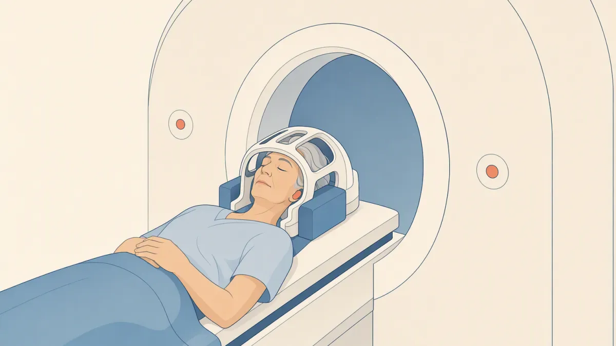
One common technique, called time-of-flight, needs no injection. An MRI image comes from hydrogen nuclei being hit with radio pulses and signaling back as they settle. Pulse the same still tissue quickly and repeatedly, and it never recovers between pulses, so it goes quiet. The scanner does exactly that to one thin slice of your head.
Blood, though, does not sit still. Fresh blood keeps flowing into that slice from outside it, fully recovered because it missed the earlier pulses. So it answers at full strength while the still tissue around it stays dim, and the arteries show up bright. Other MRA protocols use a contrast injection instead, and this explanation does not apply to them.
What it looks for
A brain MRA looks mainly for unruptured brain aneurysms — small, balloon-like bulges in an artery wall. A 2011 meta-analysis estimated that about 3 in 100 adults (Vlak 2011) carry one. Most are small and never rupture, but a rupture can be catastrophic.
- Unruptured aneurysms, with their size and location
- Narrowed arteries inside the skull (stenosis)
- Abnormal tangles of vessels (malformations)
- Very small aneurysms can be missed. A clear scan lowers the odds of a hidden aneurysm; it does not make them zero. If a specialist needs more detail, a CT angiogram or catheter angiography can follow.
- The neck arteries that feed the brain. A carotid ultrasound looks at those directly.
- The brain tissue itself. Tumors, old small strokes and structural change are read on a brain MRI, which a full-body MRI may include, depending on the protocol.
- Predicting a stroke. A normal MRA cannot exclude every future stroke or vascular event. Stroke risk is lowered by controlling blood pressure, cholesterol, diabetes and smoking — see a physical exam and blood tests, not more imaging.
Aneurysms are usually silent, but not always. A larger unruptured aneurysm can press on nearby nerves. The US National Institute of Neurological Disorders and Stroke lists the signs:
- Pain above or behind one eye
- A widened pupil, double vision or other vision change
- A drooping eyelid, or numbness on one side of the face
These need a doctor promptly, not a screening slot weeks away.
Who should consider it
This scan is not a routine check for everyone. US guidance from the American Heart Association and American Stroke Association (2015) names the groups where screening is discussed:
| Situation | What the guideline says |
|---|---|
| Two or more family members with an aneurysm or subarachnoid hemorrhage (a bleed on the brain's surface) | Screening should be offered |
| Polycystic kidney disease | Screening should be offered, particularly to those with a family history of aneurysm |
High blood pressure and smoking raise the chance of an aneurysm growing or bleeding. On their own, they are reasons to treat blood pressure and stop smoking — not established reasons to scan. It adds little in two situations:
- You have no family history and no inherited condition. The value of scanning is far less clear, and small findings can lead to years of follow-up imaging.
- You have new symptoms. That calls for a doctor's assessment first, and sudden symptoms call for 119.
What to expect on scan day
-
1Metal-safety checkYou confirm implants, devices and metal history one item at a time. Tell the team about a pacemaker, defibrillator, cochlear implant, aneurysm clip or metal fragments — some are unsafe in the magnet and others need special settings. Tell them too if you are or could be pregnant.
-
2Lie still with your head in the coilThe scanner is loud, so you are given ear protection. The MRA may run on its own or alongside a brain MRI — your coordinator confirms the session and its length when you book.
-
3Contrast only if the protocol needs itTime-of-flight MRA needs no injection. If your protocol uses gadolinium, a small IV line is placed first.
-
4AfterwardsMost people go straight back to their day. If you were given a sedative for anxiety, you need someone with you and should not drive.
Cost and coverage
The live price further down this page is the booking price at New Dawn Health; anything billed separately — such as biopsy or pathology where it applies — is confirmed when you book. What happens after that depends on where your coverage comes from:
- In Taiwan, screening is paid for by the visitor. Short-term visitors are not covered by Taiwan's National Health Insurance, so screening is self-funded.
- Reimbursement at home is a question for your insurer. Whether a scan with or without symptoms is covered depends on the plan. Ask them before you book, and ask which documents they need.
- It is usually booked with a brain MRI. Which packages include it, or offer it as an add-on, is listed below.
Results and follow-up
- A written radiologist's report. Turnaround, language and how a finding is communicated are confirmed when you book.
- A small aneurysm is usually watched, not treated. A specialist weighs its size, location and your risk factors, and often sets a repeat-imaging interval.
- A finding is not an emergency by itself — unless new symptoms appear, which call for urgent care.
- A clear scan becomes a baseline. It does not exclude every future stroke or vascular event.